Calcium »
PDB 1m9i-1mr8 »
1mdu »
Calcium in PDB 1mdu: Crystal Structure of the Chicken Actin Trimer Complexed with Human Gelsolin Segment 1 (Gs-1)
Protein crystallography data
The structure of Crystal Structure of the Chicken Actin Trimer Complexed with Human Gelsolin Segment 1 (Gs-1), PDB code: 1mdu
was solved by
J.F.Dawson,
E.P.Sablin,
J.A.Spudich,
R.J.Fletterick,
with X-Ray Crystallography technique. A brief refinement statistics is given in the table below:
| Resolution Low / High (Å) | 25.00 / 2.20 |
| Space group | P 1 21 1 |
| Cell size a, b, c (Å), α, β, γ (°) | 67.187, 75.942, 96.747, 90.00, 91.87, 90.00 |
| R / Rfree (%) | 19.3 / 23.3 |
Calcium Binding Sites:
The binding sites of Calcium atom in the Crystal Structure of the Chicken Actin Trimer Complexed with Human Gelsolin Segment 1 (Gs-1)
(pdb code 1mdu). This binding sites where shown within
5.0 Angstroms radius around Calcium atom.
In total 7 binding sites of Calcium where determined in the Crystal Structure of the Chicken Actin Trimer Complexed with Human Gelsolin Segment 1 (Gs-1), PDB code: 1mdu:
Jump to Calcium binding site number: 1; 2; 3; 4; 5; 6; 7;
In total 7 binding sites of Calcium where determined in the Crystal Structure of the Chicken Actin Trimer Complexed with Human Gelsolin Segment 1 (Gs-1), PDB code: 1mdu:
Jump to Calcium binding site number: 1; 2; 3; 4; 5; 6; 7;
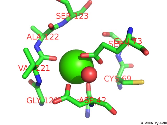
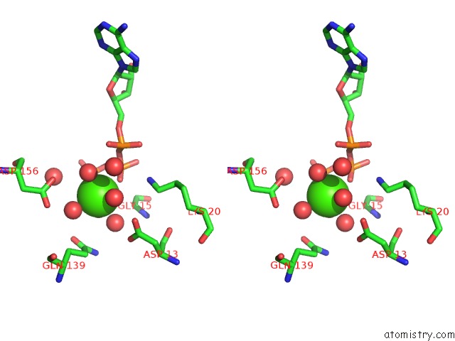
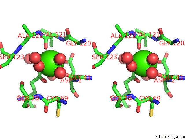
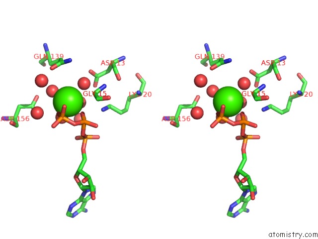
Calcium binding site 1 out of 7 in 1mdu
Go back to
Calcium binding site 1 out
of 7 in the Crystal Structure of the Chicken Actin Trimer Complexed with Human Gelsolin Segment 1 (Gs-1)
Mono view
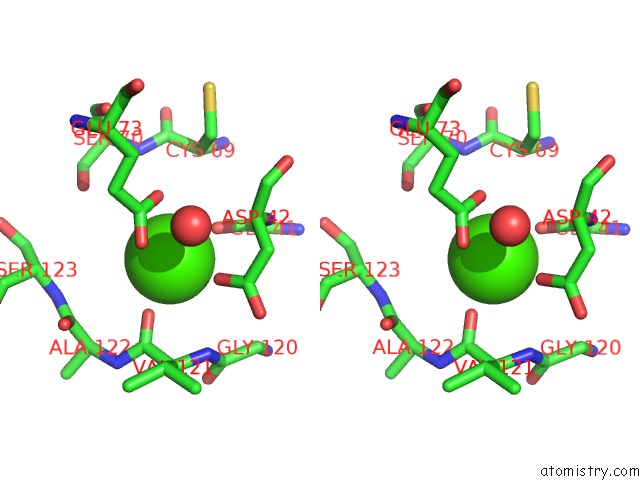
Stereo pair view
Mono view
Stereo pair view
A full contact list of Calcium with other atoms in the Ca binding
site number 1 of Crystal Structure of the Chicken Actin Trimer Complexed with Human Gelsolin Segment 1 (Gs-1) within 5.0Å range:
|
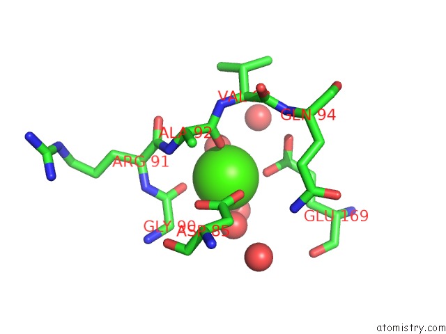
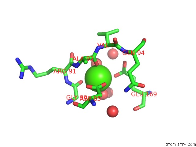
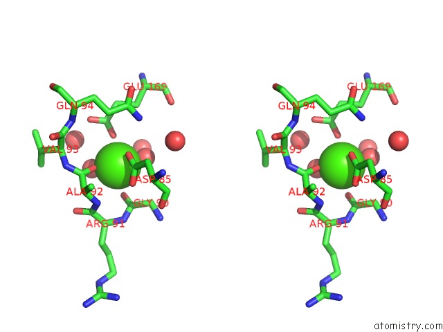
Calcium binding site 2 out of 7 in 1mdu
Go back to
Calcium binding site 2 out
of 7 in the Crystal Structure of the Chicken Actin Trimer Complexed with Human Gelsolin Segment 1 (Gs-1)
Mono view

Stereo pair view
Mono view
Stereo pair view
A full contact list of Calcium with other atoms in the Ca binding
site number 2 of Crystal Structure of the Chicken Actin Trimer Complexed with Human Gelsolin Segment 1 (Gs-1) within 5.0Å range:
|
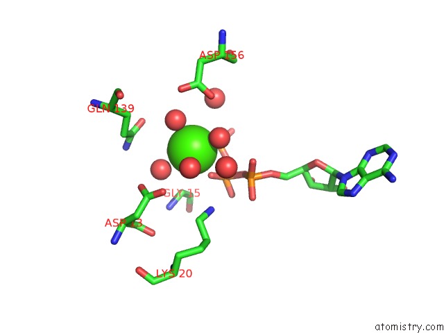
Calcium binding site 3 out of 7 in 1mdu
Go back to
Calcium binding site 3 out
of 7 in the Crystal Structure of the Chicken Actin Trimer Complexed with Human Gelsolin Segment 1 (Gs-1)
Mono view
Stereo pair view
Mono view
Stereo pair view
A full contact list of Calcium with other atoms in the Ca binding
site number 3 of Crystal Structure of the Chicken Actin Trimer Complexed with Human Gelsolin Segment 1 (Gs-1) within 5.0Å range:
|
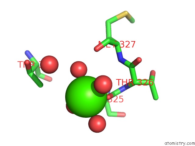
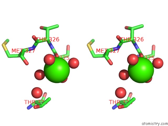
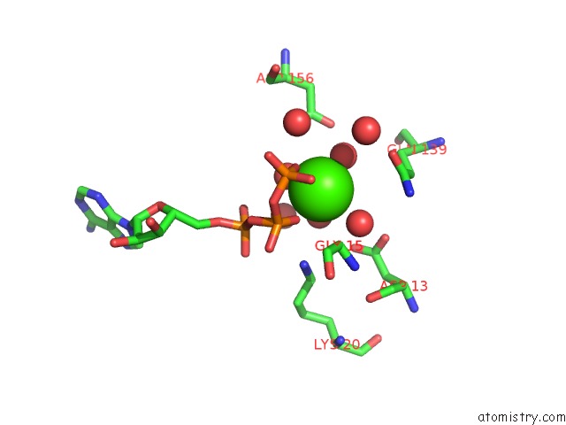
Calcium binding site 4 out of 7 in 1mdu
Go back to
Calcium binding site 4 out
of 7 in the Crystal Structure of the Chicken Actin Trimer Complexed with Human Gelsolin Segment 1 (Gs-1)
Mono view
Stereo pair view
Mono view
Stereo pair view
A full contact list of Calcium with other atoms in the Ca binding
site number 4 of Crystal Structure of the Chicken Actin Trimer Complexed with Human Gelsolin Segment 1 (Gs-1) within 5.0Å range:
|
Calcium binding site 5 out of 7 in 1mdu
Go back to
Calcium binding site 5 out
of 7 in the Crystal Structure of the Chicken Actin Trimer Complexed with Human Gelsolin Segment 1 (Gs-1)
Mono view
Stereo pair view
Mono view
Stereo pair view
A full contact list of Calcium with other atoms in the Ca binding
site number 5 of Crystal Structure of the Chicken Actin Trimer Complexed with Human Gelsolin Segment 1 (Gs-1) within 5.0Å range:
|
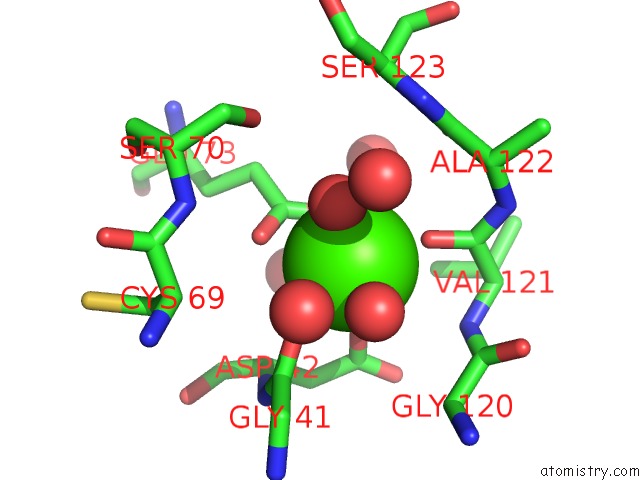
Calcium binding site 6 out of 7 in 1mdu
Go back to
Calcium binding site 6 out
of 7 in the Crystal Structure of the Chicken Actin Trimer Complexed with Human Gelsolin Segment 1 (Gs-1)
Mono view
Stereo pair view
Mono view
Stereo pair view
A full contact list of Calcium with other atoms in the Ca binding
site number 6 of Crystal Structure of the Chicken Actin Trimer Complexed with Human Gelsolin Segment 1 (Gs-1) within 5.0Å range:
|
Calcium binding site 7 out of 7 in 1mdu
Go back to
Calcium binding site 7 out
of 7 in the Crystal Structure of the Chicken Actin Trimer Complexed with Human Gelsolin Segment 1 (Gs-1)
Mono view
Stereo pair view
Mono view
Stereo pair view
A full contact list of Calcium with other atoms in the Ca binding
site number 7 of Crystal Structure of the Chicken Actin Trimer Complexed with Human Gelsolin Segment 1 (Gs-1) within 5.0Å range:
|
Reference:
J.F.Dawson,
E.P.Sablin,
J.A.Spudich,
R.J.Fletterick.
Structure of An F-Actin Trimer Disrupted By Gelsolin and Implications For the Mechanism of Severing J.Biol.Chem. V. 278 1229 2003.
ISSN: ISSN 0021-9258
PubMed: 12356759
DOI: 10.1074/JBC.M209160200
Page generated: Thu Jul 11 12:29:57 2024
ISSN: ISSN 0021-9258
PubMed: 12356759
DOI: 10.1074/JBC.M209160200
Last articles
Zn in 9JYWZn in 9IR4
Zn in 9IR3
Zn in 9GMX
Zn in 9GMW
Zn in 9JEJ
Zn in 9ERF
Zn in 9ERE
Zn in 9EGV
Zn in 9EGW