Calcium »
PDB 1m9i-1mr8 »
1mdw »
Calcium in PDB 1mdw: Crystal Structure of Calcium-Bound Protease Core of Calpain II Reveals the Basis For Intrinsic Inactivation
Enzymatic activity of Crystal Structure of Calcium-Bound Protease Core of Calpain II Reveals the Basis For Intrinsic Inactivation
All present enzymatic activity of Crystal Structure of Calcium-Bound Protease Core of Calpain II Reveals the Basis For Intrinsic Inactivation:
3.4.22.17;
3.4.22.17;
Protein crystallography data
The structure of Crystal Structure of Calcium-Bound Protease Core of Calpain II Reveals the Basis For Intrinsic Inactivation, PDB code: 1mdw
was solved by
T.Moldoveanu,
C.M.Hosfield,
D.Lim,
Z.Jia,
P.L.Davies,
with X-Ray Crystallography technique. A brief refinement statistics is given in the table below:
| Resolution Low / High (Å) | 20.00 / 1.95 |
| Space group | P 1 21 1 |
| Cell size a, b, c (Å), α, β, γ (°) | 62.600, 80.600, 75.300, 90.00, 103.90, 90.00 |
| R / Rfree (%) | 20.7 / 24.3 |
Calcium Binding Sites:
The binding sites of Calcium atom in the Crystal Structure of Calcium-Bound Protease Core of Calpain II Reveals the Basis For Intrinsic Inactivation
(pdb code 1mdw). This binding sites where shown within
5.0 Angstroms radius around Calcium atom.
In total 4 binding sites of Calcium where determined in the Crystal Structure of Calcium-Bound Protease Core of Calpain II Reveals the Basis For Intrinsic Inactivation, PDB code: 1mdw:
Jump to Calcium binding site number: 1; 2; 3; 4;
In total 4 binding sites of Calcium where determined in the Crystal Structure of Calcium-Bound Protease Core of Calpain II Reveals the Basis For Intrinsic Inactivation, PDB code: 1mdw:
Jump to Calcium binding site number: 1; 2; 3; 4;
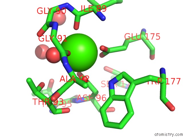
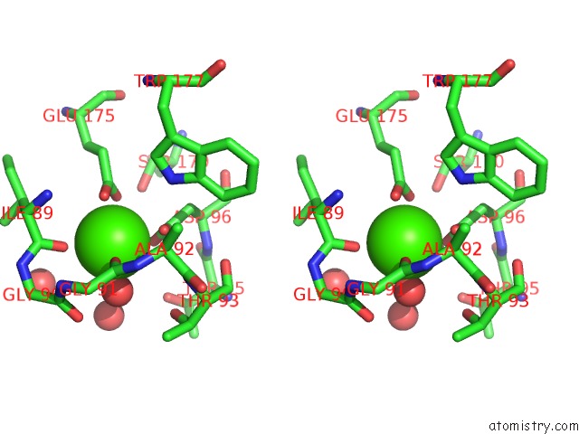
Calcium binding site 1 out of 4 in 1mdw
Go back to
Calcium binding site 1 out
of 4 in the Crystal Structure of Calcium-Bound Protease Core of Calpain II Reveals the Basis For Intrinsic Inactivation
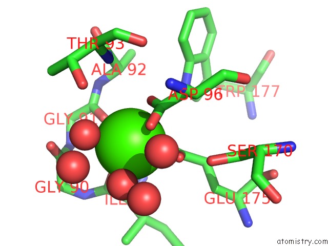
Mono view
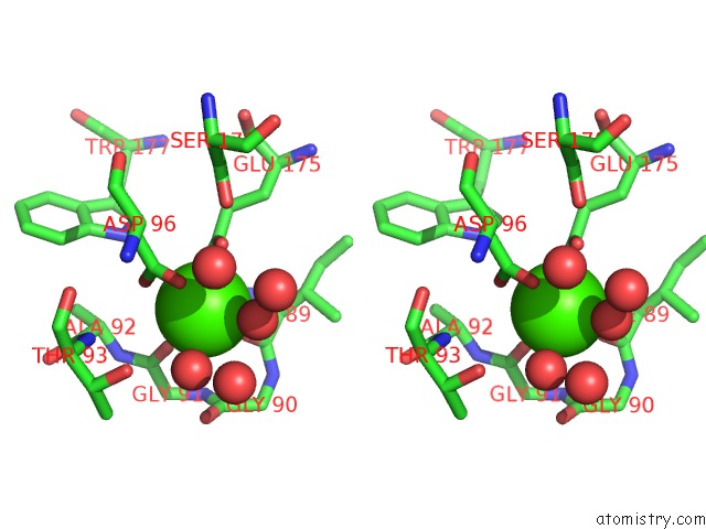
Stereo pair view
Mono view
Stereo pair view
A full contact list of Calcium with other atoms in the Ca binding
site number 1 of Crystal Structure of Calcium-Bound Protease Core of Calpain II Reveals the Basis For Intrinsic Inactivation within 5.0Å range:
|
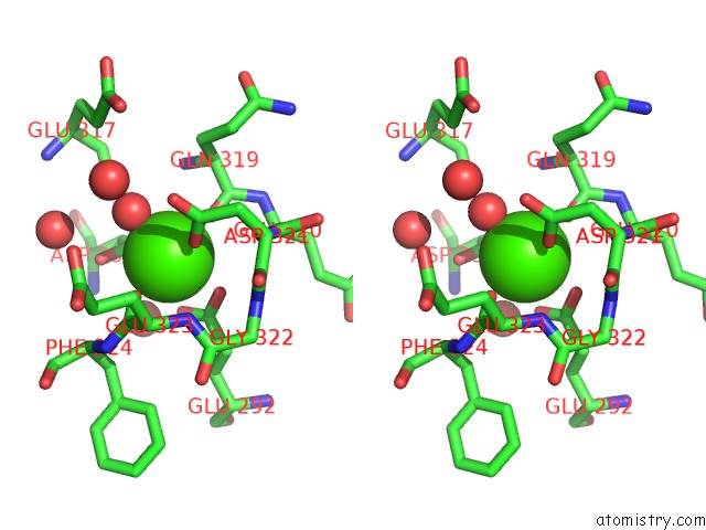
Calcium binding site 2 out of 4 in 1mdw
Go back to
Calcium binding site 2 out
of 4 in the Crystal Structure of Calcium-Bound Protease Core of Calpain II Reveals the Basis For Intrinsic Inactivation
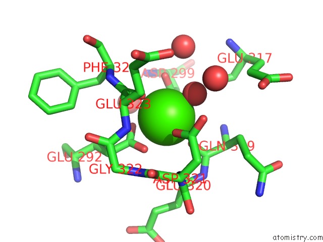
Mono view
Stereo pair view
Mono view
Stereo pair view
A full contact list of Calcium with other atoms in the Ca binding
site number 2 of Crystal Structure of Calcium-Bound Protease Core of Calpain II Reveals the Basis For Intrinsic Inactivation within 5.0Å range:
|
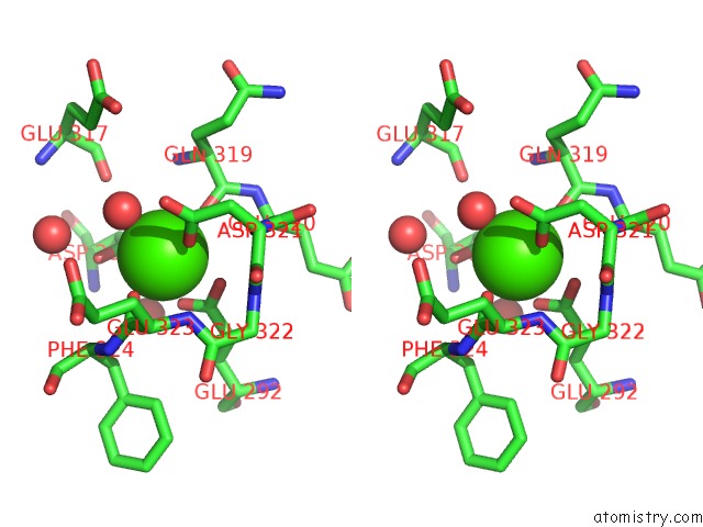
Calcium binding site 3 out of 4 in 1mdw
Go back to
Calcium binding site 3 out
of 4 in the Crystal Structure of Calcium-Bound Protease Core of Calpain II Reveals the Basis For Intrinsic Inactivation
Mono view
Stereo pair view
Mono view
Stereo pair view
A full contact list of Calcium with other atoms in the Ca binding
site number 3 of Crystal Structure of Calcium-Bound Protease Core of Calpain II Reveals the Basis For Intrinsic Inactivation within 5.0Å range:
|
Calcium binding site 4 out of 4 in 1mdw
Go back to
Calcium binding site 4 out
of 4 in the Crystal Structure of Calcium-Bound Protease Core of Calpain II Reveals the Basis For Intrinsic Inactivation
Mono view
Stereo pair view
Mono view
Stereo pair view
A full contact list of Calcium with other atoms in the Ca binding
site number 4 of Crystal Structure of Calcium-Bound Protease Core of Calpain II Reveals the Basis For Intrinsic Inactivation within 5.0Å range:
|
Reference:
T.Moldoveanu,
C.M.Hosfield,
D.Lim,
Z.Jia,
P.L.Davies.
Calpain Silencing By A Reversible Intrinsic Mechanism. Nat.Struct.Biol. V. 10 371 2003.
ISSN: ISSN 1072-8368
PubMed: 12665854
DOI: 10.1038/NSB917
Page generated: Thu Jul 11 12:30:02 2024
ISSN: ISSN 1072-8368
PubMed: 12665854
DOI: 10.1038/NSB917
Last articles
Zn in 9J0NZn in 9J0O
Zn in 9J0P
Zn in 9FJX
Zn in 9EKB
Zn in 9C0F
Zn in 9CAH
Zn in 9CH0
Zn in 9CH3
Zn in 9CH1