Calcium »
PDB 1m9i-1mr8 »
1mks »
Calcium in PDB 1mks: Carboxylic Ester Hydrolase, Trigonal Form of the Triple Mutant
Enzymatic activity of Carboxylic Ester Hydrolase, Trigonal Form of the Triple Mutant
All present enzymatic activity of Carboxylic Ester Hydrolase, Trigonal Form of the Triple Mutant:
3.1.1.4;
3.1.1.4;
Protein crystallography data
The structure of Carboxylic Ester Hydrolase, Trigonal Form of the Triple Mutant, PDB code: 1mks
was solved by
M.Sundaralingam,
with X-Ray Crystallography technique. A brief refinement statistics is given in the table below:
| Resolution Low / High (Å) | 8.00 / 1.90 |
| Space group | P 31 2 1 |
| Cell size a, b, c (Å), α, β, γ (°) | 47.080, 47.080, 102.370, 90.00, 90.00, 120.00 |
| R / Rfree (%) | 18.6 / n/a |
Calcium Binding Sites:
The binding sites of Calcium atom in the Carboxylic Ester Hydrolase, Trigonal Form of the Triple Mutant
(pdb code 1mks). This binding sites where shown within
5.0 Angstroms radius around Calcium atom.
In total only one binding site of Calcium was determined in the Carboxylic Ester Hydrolase, Trigonal Form of the Triple Mutant, PDB code: 1mks:
In total only one binding site of Calcium was determined in the Carboxylic Ester Hydrolase, Trigonal Form of the Triple Mutant, PDB code: 1mks:
Calcium binding site 1 out of 1 in 1mks
Go back to
Calcium binding site 1 out
of 1 in the Carboxylic Ester Hydrolase, Trigonal Form of the Triple Mutant
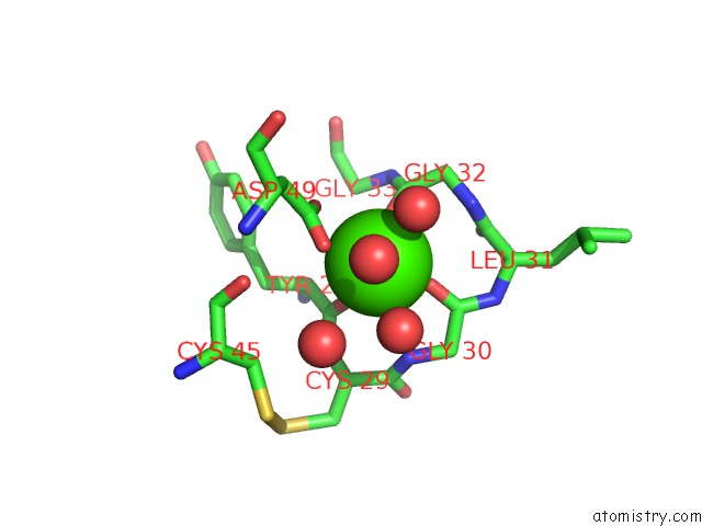
Mono view
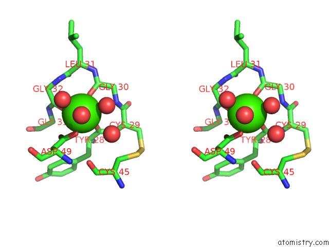
Stereo pair view
Mono view
Stereo pair view
A full contact list of Calcium with other atoms in the Ca binding
site number 1 of Carboxylic Ester Hydrolase, Trigonal Form of the Triple Mutant within 5.0Å range:
|
Reference:
K.Sekar,
B.Z.Yu,
J.Rogers,
J.Lutton,
X.Liu,
X.Chen,
M.D.Tsai,
M.K.Jain,
M.Sundaralingam.
Phospholipase A2 Engineering. Structural and Functional Roles of the Highly Conserved Active Site Residue Aspartate-99. Biochemistry V. 36 3104 1997.
ISSN: ISSN 0006-2960
PubMed: 9115986
DOI: 10.1021/BI961576X
Page generated: Thu Jul 11 12:33:05 2024
ISSN: ISSN 0006-2960
PubMed: 9115986
DOI: 10.1021/BI961576X
Last articles
Zn in 9J0NZn in 9J0O
Zn in 9J0P
Zn in 9FJX
Zn in 9EKB
Zn in 9C0F
Zn in 9CAH
Zn in 9CH0
Zn in 9CH3
Zn in 9CH1