Calcium »
PDB 1mts-1n7d »
1mwn »
Calcium in PDB 1mwn: Solution uc(Nmr) Structure of S100B Bound to the High-Affinity Target Peptide Trtk-12
Calcium Binding Sites:
The binding sites of Calcium atom in the Solution uc(Nmr) Structure of S100B Bound to the High-Affinity Target Peptide Trtk-12
(pdb code 1mwn). This binding sites where shown within
5.0 Angstroms radius around Calcium atom.
In total 4 binding sites of Calcium where determined in the Solution uc(Nmr) Structure of S100B Bound to the High-Affinity Target Peptide Trtk-12, PDB code: 1mwn:
Jump to Calcium binding site number: 1; 2; 3; 4;
In total 4 binding sites of Calcium where determined in the Solution uc(Nmr) Structure of S100B Bound to the High-Affinity Target Peptide Trtk-12, PDB code: 1mwn:
Jump to Calcium binding site number: 1; 2; 3; 4;
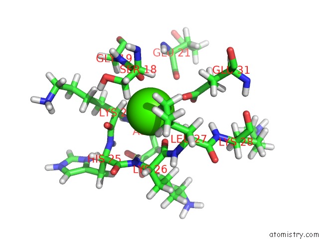
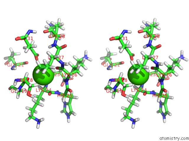
Calcium binding site 1 out of 4 in 1mwn
Go back to
Calcium binding site 1 out
of 4 in the Solution uc(Nmr) Structure of S100B Bound to the High-Affinity Target Peptide Trtk-12
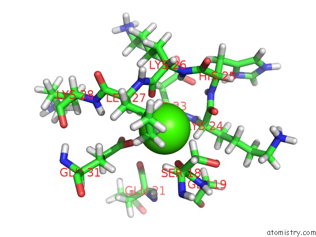
Mono view
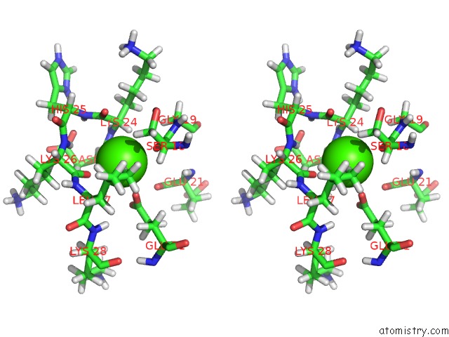
Stereo pair view
Mono view
Stereo pair view
A full contact list of Calcium with other atoms in the Ca binding
site number 1 of Solution uc(Nmr) Structure of S100B Bound to the High-Affinity Target Peptide Trtk-12 within 5.0Å range:
|
Calcium binding site 2 out of 4 in 1mwn
Go back to
Calcium binding site 2 out
of 4 in the Solution uc(Nmr) Structure of S100B Bound to the High-Affinity Target Peptide Trtk-12
Mono view
Stereo pair view
Mono view
Stereo pair view
A full contact list of Calcium with other atoms in the Ca binding
site number 2 of Solution uc(Nmr) Structure of S100B Bound to the High-Affinity Target Peptide Trtk-12 within 5.0Å range:
|
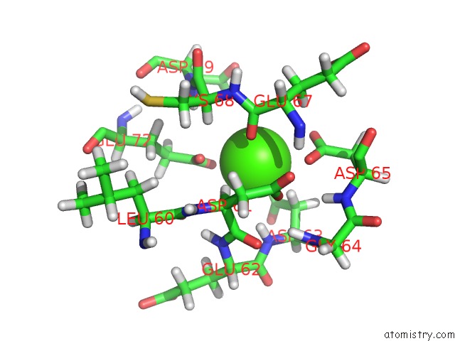
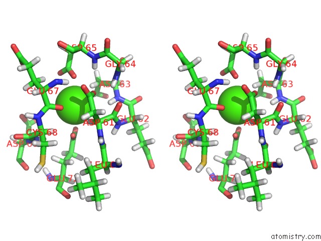
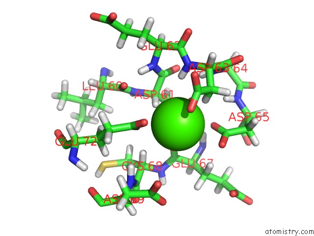
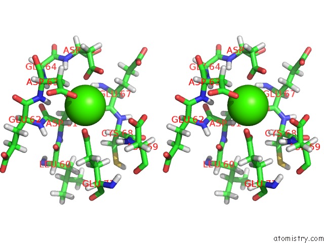
Calcium binding site 3 out of 4 in 1mwn
Go back to
Calcium binding site 3 out
of 4 in the Solution uc(Nmr) Structure of S100B Bound to the High-Affinity Target Peptide Trtk-12
Mono view
Stereo pair view
Mono view
Stereo pair view
A full contact list of Calcium with other atoms in the Ca binding
site number 3 of Solution uc(Nmr) Structure of S100B Bound to the High-Affinity Target Peptide Trtk-12 within 5.0Å range:
|
Calcium binding site 4 out of 4 in 1mwn
Go back to
Calcium binding site 4 out
of 4 in the Solution uc(Nmr) Structure of S100B Bound to the High-Affinity Target Peptide Trtk-12
Mono view
Stereo pair view
Mono view
Stereo pair view
A full contact list of Calcium with other atoms in the Ca binding
site number 4 of Solution uc(Nmr) Structure of S100B Bound to the High-Affinity Target Peptide Trtk-12 within 5.0Å range:
|
Reference:
K.G.Inman,
R.Yang,
R.R.Rustandi,
K.E.Miller,
D.M.Baldisseri,
D.J.Weber.
Solution uc(Nmr) Structure of S100B Bound to the High-Affinity Target Peptide Trtk-12 J.Mol.Biol. V. 324 1003 2002.
ISSN: ISSN 0022-2836
PubMed: 12470955
DOI: 10.1016/S0022-2836(02)01152-X
Page generated: Thu Jul 11 12:38:19 2024
ISSN: ISSN 0022-2836
PubMed: 12470955
DOI: 10.1016/S0022-2836(02)01152-X
Last articles
Zn in 9MJ5Zn in 9HNW
Zn in 9G0L
Zn in 9FNE
Zn in 9DZN
Zn in 9E0I
Zn in 9D32
Zn in 9DAK
Zn in 8ZXC
Zn in 8ZUF