Calcium »
PDB 1mts-1n7d »
1n41 »
Calcium in PDB 1n41: Crystal Structure of Annexin V K27E Mutant
Protein crystallography data
The structure of Crystal Structure of Annexin V K27E Mutant, PDB code: 1n41
was solved by
Y.D.Mo,
B.Campos,
T.R.Mealy,
L.Commodore,
J.F.Head,
J.R.Dedman,
B.A.Seaton,
with X-Ray Crystallography technique. A brief refinement statistics is given in the table below:
| Resolution Low / High (Å) | 50.00 / 2.10 |
| Space group | H 3 |
| Cell size a, b, c (Å), α, β, γ (°) | 157.596, 157.596, 37.059, 90.00, 90.00, 120.00 |
| R / Rfree (%) | 18.7 / 22.3 |
Calcium Binding Sites:
The binding sites of Calcium atom in the Crystal Structure of Annexin V K27E Mutant
(pdb code 1n41). This binding sites where shown within
5.0 Angstroms radius around Calcium atom.
In total 5 binding sites of Calcium where determined in the Crystal Structure of Annexin V K27E Mutant, PDB code: 1n41:
Jump to Calcium binding site number: 1; 2; 3; 4; 5;
In total 5 binding sites of Calcium where determined in the Crystal Structure of Annexin V K27E Mutant, PDB code: 1n41:
Jump to Calcium binding site number: 1; 2; 3; 4; 5;
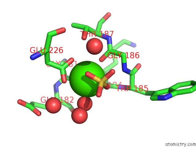
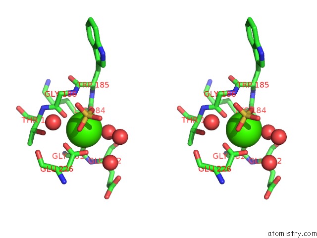
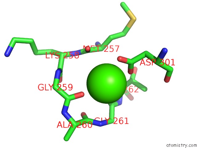
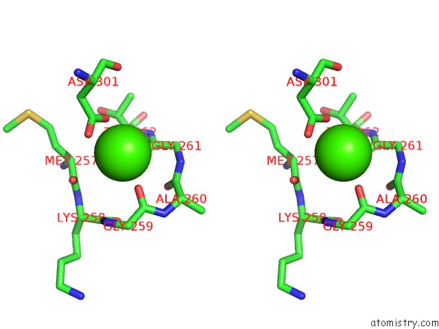
Calcium binding site 1 out of 5 in 1n41
Go back to
Calcium binding site 1 out
of 5 in the Crystal Structure of Annexin V K27E Mutant
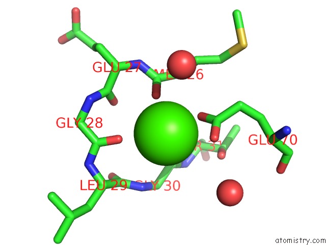
Mono view
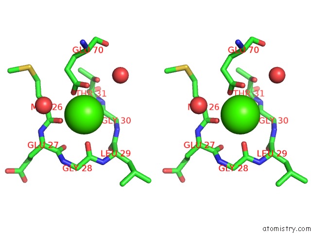
Stereo pair view
Mono view
Stereo pair view
A full contact list of Calcium with other atoms in the Ca binding
site number 1 of Crystal Structure of Annexin V K27E Mutant within 5.0Å range:
|
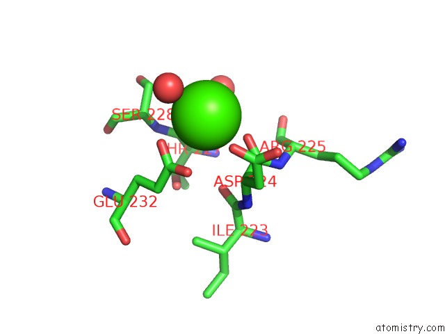
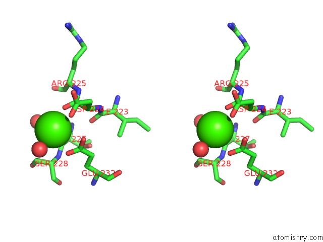
Calcium binding site 2 out of 5 in 1n41
Go back to
Calcium binding site 2 out
of 5 in the Crystal Structure of Annexin V K27E Mutant
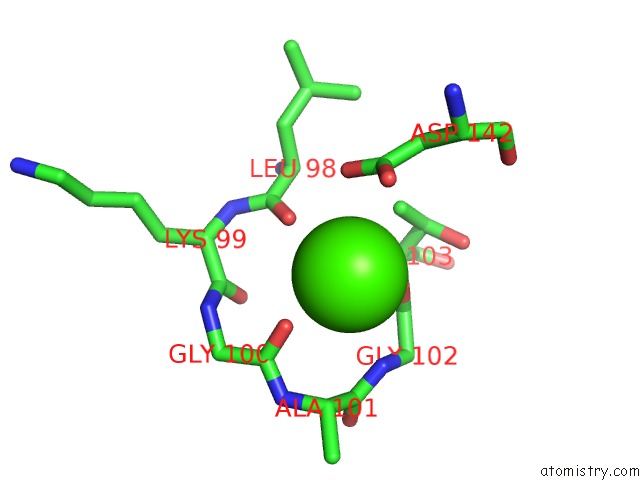
Mono view
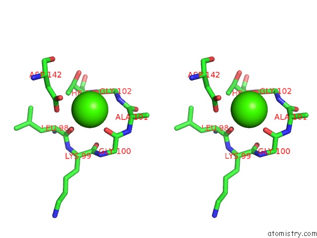
Stereo pair view
Mono view
Stereo pair view
A full contact list of Calcium with other atoms in the Ca binding
site number 2 of Crystal Structure of Annexin V K27E Mutant within 5.0Å range:
|
Calcium binding site 3 out of 5 in 1n41
Go back to
Calcium binding site 3 out
of 5 in the Crystal Structure of Annexin V K27E Mutant
Mono view
Stereo pair view
Mono view
Stereo pair view
A full contact list of Calcium with other atoms in the Ca binding
site number 3 of Crystal Structure of Annexin V K27E Mutant within 5.0Å range:
|
Calcium binding site 4 out of 5 in 1n41
Go back to
Calcium binding site 4 out
of 5 in the Crystal Structure of Annexin V K27E Mutant
Mono view
Stereo pair view
Mono view
Stereo pair view
A full contact list of Calcium with other atoms in the Ca binding
site number 4 of Crystal Structure of Annexin V K27E Mutant within 5.0Å range:
|
Calcium binding site 5 out of 5 in 1n41
Go back to
Calcium binding site 5 out
of 5 in the Crystal Structure of Annexin V K27E Mutant
Mono view
Stereo pair view
Mono view
Stereo pair view
A full contact list of Calcium with other atoms in the Ca binding
site number 5 of Crystal Structure of Annexin V K27E Mutant within 5.0Å range:
|
Reference:
Y.D.Mo,
B.Campos,
T.R.Mealy,
L.Commodore,
J.F.Head,
J.R.Dedman,
B.A.Seaton.
Interfacial Basic Cluster in Annexin V Couples Phospholipid Binding and Trimer Formation on Membrane Surfaces J.Biol.Chem. V. 278 2437 2003.
ISSN: ISSN 0021-9258
PubMed: 12401794
DOI: 10.1074/JBC.M210286200
Page generated: Mon Jul 7 17:26:51 2025
ISSN: ISSN 0021-9258
PubMed: 12401794
DOI: 10.1074/JBC.M210286200
Last articles
F in 7PRXF in 7PPH
F in 7PRM
F in 7PQV
F in 7PQS
F in 7PMM
F in 7POL
F in 7POR
F in 7PNS
F in 7P81