Calcium »
PDB 1mts-1n7d »
1n44 »
Calcium in PDB 1n44: Crystal Structure of Annexin V R23E Mutant
Protein crystallography data
The structure of Crystal Structure of Annexin V R23E Mutant, PDB code: 1n44
was solved by
Y.D.Mo,
B.Campos,
T.R.Mealy,
L.Commodore,
J.F.Head,
J.R.Dedman,
B.A.Seaton,
with X-Ray Crystallography technique. A brief refinement statistics is given in the table below:
| Resolution Low / High (Å) | 50.00 / 3.00 |
| Space group | H 3 |
| Cell size a, b, c (Å), α, β, γ (°) | 157.757, 157.757, 36.761, 90.00, 90.00, 120.00 |
| R / Rfree (%) | 16.3 / 24.1 |
Calcium Binding Sites:
The binding sites of Calcium atom in the Crystal Structure of Annexin V R23E Mutant
(pdb code 1n44). This binding sites where shown within
5.0 Angstroms radius around Calcium atom.
In total 2 binding sites of Calcium where determined in the Crystal Structure of Annexin V R23E Mutant, PDB code: 1n44:
Jump to Calcium binding site number: 1; 2;
In total 2 binding sites of Calcium where determined in the Crystal Structure of Annexin V R23E Mutant, PDB code: 1n44:
Jump to Calcium binding site number: 1; 2;
Calcium binding site 1 out of 2 in 1n44
Go back to
Calcium binding site 1 out
of 2 in the Crystal Structure of Annexin V R23E Mutant
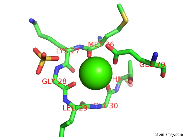
Mono view
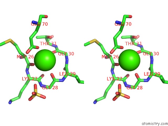
Stereo pair view
Mono view
Stereo pair view
A full contact list of Calcium with other atoms in the Ca binding
site number 1 of Crystal Structure of Annexin V R23E Mutant within 5.0Å range:
|
Calcium binding site 2 out of 2 in 1n44
Go back to
Calcium binding site 2 out
of 2 in the Crystal Structure of Annexin V R23E Mutant
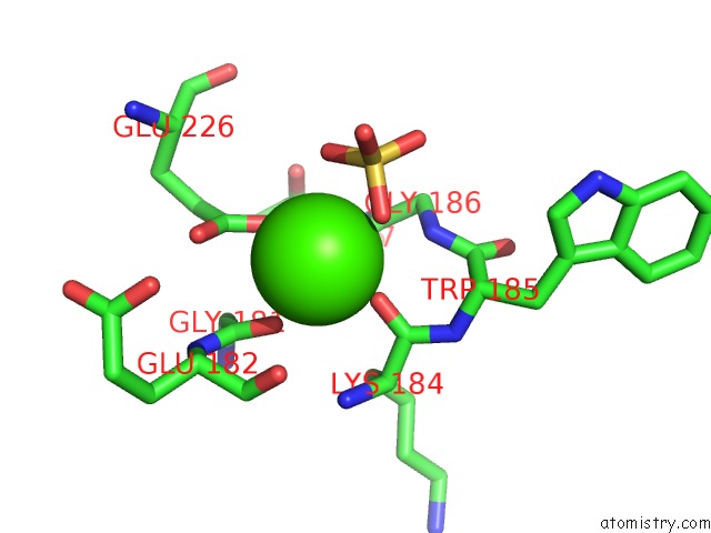
Mono view
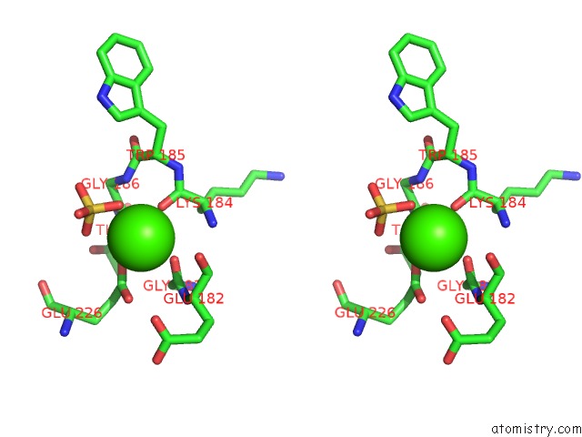
Stereo pair view
Mono view
Stereo pair view
A full contact list of Calcium with other atoms in the Ca binding
site number 2 of Crystal Structure of Annexin V R23E Mutant within 5.0Å range:
|
Reference:
Y.D.Mo,
B.Campos,
T.R.Mealy,
L.Commodore,
J.F.Head,
J.R.Dedman,
B.A.Seaton.
Interfacial Basic Cluster in Anexin V Couples Phospholipid Binding and Trimer Formation on Membrane Surfaces J.Biol.Chem. V. 278 2437 2003.
ISSN: ISSN 0021-9258
PubMed: 12401794
DOI: 10.1074/JBC.M210286200
Page generated: Thu Jul 11 12:43:25 2024
ISSN: ISSN 0021-9258
PubMed: 12401794
DOI: 10.1074/JBC.M210286200
Last articles
Zn in 9MJ5Zn in 9HNW
Zn in 9G0L
Zn in 9FNE
Zn in 9DZN
Zn in 9E0I
Zn in 9D32
Zn in 9DAK
Zn in 8ZXC
Zn in 8ZUF