Calcium »
PDB 1n7s-1nm9 »
1n7u »
Calcium in PDB 1n7u: The Receptor-Binding Protein P2 of Bacteriophage PRD1: Crystal Form I
Protein crystallography data
The structure of The Receptor-Binding Protein P2 of Bacteriophage PRD1: Crystal Form I, PDB code: 1n7u
was solved by
L.Xu,
S.D.Benson,
S.J.Butcher,
D.H.Bamford,
R.M.Burnett,
with X-Ray Crystallography technique. A brief refinement statistics is given in the table below:
| Resolution Low / High (Å) | 43.22 / 2.40 |
| Space group | P 2 2 21 |
| Cell size a, b, c (Å), α, β, γ (°) | 138.200, 46.500, 136.500, 90.00, 90.00, 90.00 |
| R / Rfree (%) | 22.4 / 24.8 |
Calcium Binding Sites:
The binding sites of Calcium atom in the The Receptor-Binding Protein P2 of Bacteriophage PRD1: Crystal Form I
(pdb code 1n7u). This binding sites where shown within
5.0 Angstroms radius around Calcium atom.
In total only one binding site of Calcium was determined in the The Receptor-Binding Protein P2 of Bacteriophage PRD1: Crystal Form I, PDB code: 1n7u:
In total only one binding site of Calcium was determined in the The Receptor-Binding Protein P2 of Bacteriophage PRD1: Crystal Form I, PDB code: 1n7u:
Calcium binding site 1 out of 1 in 1n7u
Go back to
Calcium binding site 1 out
of 1 in the The Receptor-Binding Protein P2 of Bacteriophage PRD1: Crystal Form I
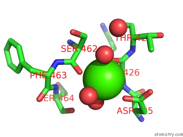
Mono view
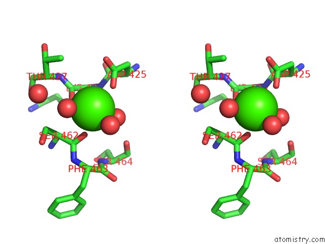
Stereo pair view
Mono view
Stereo pair view
A full contact list of Calcium with other atoms in the Ca binding
site number 1 of The Receptor-Binding Protein P2 of Bacteriophage PRD1: Crystal Form I within 5.0Å range:
|
Reference:
L.Xu,
S.D.Benson,
S.J.Butcher,
D.H.Bamford,
R.M.Burnett.
The Receptor Binding Protein P2 of PRD1, A Virus Targeting Antibiotic-Resistant Bacteria, Has A Novel Fold Suggesting Multiple Functions. Structure V. 11 309 2003.
ISSN: ISSN 0969-2126
PubMed: 12623018
DOI: 10.1016/S0969-2126(03)00023-6
Page generated: Mon Jul 7 17:29:33 2025
ISSN: ISSN 0969-2126
PubMed: 12623018
DOI: 10.1016/S0969-2126(03)00023-6
Last articles
Ca in 7LTDCa in 7LSJ
Ca in 7LSU
Ca in 7LST
Ca in 7LSR
Ca in 7LSK
Ca in 7LRY
Ca in 7LSA
Ca in 7LRX
Ca in 7LRM