Calcium »
PDB 1n7s-1nm9 »
1n86 »
Calcium in PDB 1n86: Crystal Structure of Human D-Dimer From Cross-Linked Fibrin Complexed with Gpr and Ghrpldk Peptide Ligands.
Protein crystallography data
The structure of Crystal Structure of Human D-Dimer From Cross-Linked Fibrin Complexed with Gpr and Ghrpldk Peptide Ligands., PDB code: 1n86
was solved by
Z.Yang,
L.Pandi,
R.F.Doolittle,
with X-Ray Crystallography technique. A brief refinement statistics is given in the table below:
| Resolution Low / High (Å) | 20.00 / 3.20 |
| Space group | P 21 21 21 |
| Cell size a, b, c (Å), α, β, γ (°) | 52.328, 130.093, 298.313, 90.00, 90.00, 90.00 |
| R / Rfree (%) | 22.6 / 28.9 |
Calcium Binding Sites:
The binding sites of Calcium atom in the Crystal Structure of Human D-Dimer From Cross-Linked Fibrin Complexed with Gpr and Ghrpldk Peptide Ligands.
(pdb code 1n86). This binding sites where shown within
5.0 Angstroms radius around Calcium atom.
In total 4 binding sites of Calcium where determined in the Crystal Structure of Human D-Dimer From Cross-Linked Fibrin Complexed with Gpr and Ghrpldk Peptide Ligands., PDB code: 1n86:
Jump to Calcium binding site number: 1; 2; 3; 4;
In total 4 binding sites of Calcium where determined in the Crystal Structure of Human D-Dimer From Cross-Linked Fibrin Complexed with Gpr and Ghrpldk Peptide Ligands., PDB code: 1n86:
Jump to Calcium binding site number: 1; 2; 3; 4;
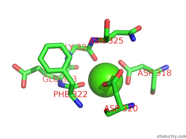
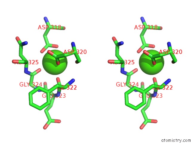
Calcium binding site 1 out of 4 in 1n86
Go back to
Calcium binding site 1 out
of 4 in the Crystal Structure of Human D-Dimer From Cross-Linked Fibrin Complexed with Gpr and Ghrpldk Peptide Ligands.
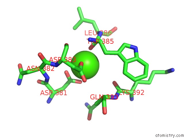
Mono view
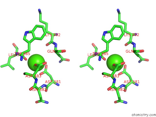
Stereo pair view
Mono view
Stereo pair view
A full contact list of Calcium with other atoms in the Ca binding
site number 1 of Crystal Structure of Human D-Dimer From Cross-Linked Fibrin Complexed with Gpr and Ghrpldk Peptide Ligands. within 5.0Å range:
|
Calcium binding site 2 out of 4 in 1n86
Go back to
Calcium binding site 2 out
of 4 in the Crystal Structure of Human D-Dimer From Cross-Linked Fibrin Complexed with Gpr and Ghrpldk Peptide Ligands.
Mono view
Stereo pair view
Mono view
Stereo pair view
A full contact list of Calcium with other atoms in the Ca binding
site number 2 of Crystal Structure of Human D-Dimer From Cross-Linked Fibrin Complexed with Gpr and Ghrpldk Peptide Ligands. within 5.0Å range:
|
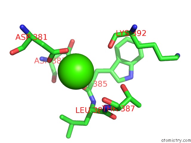
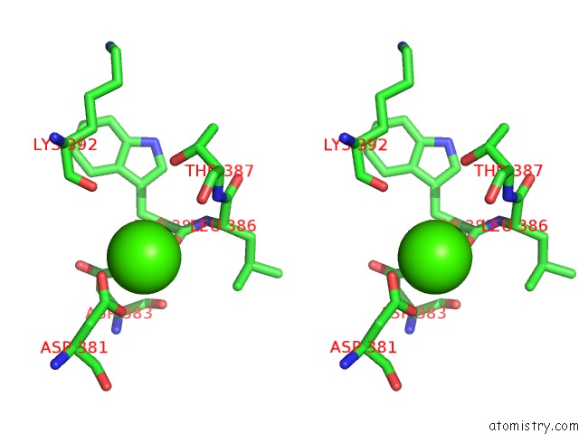
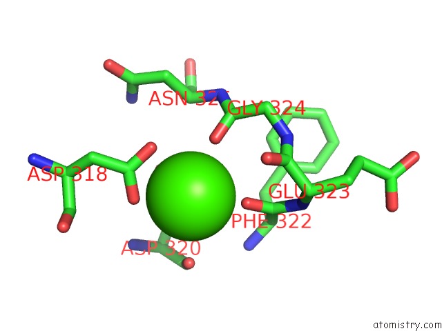
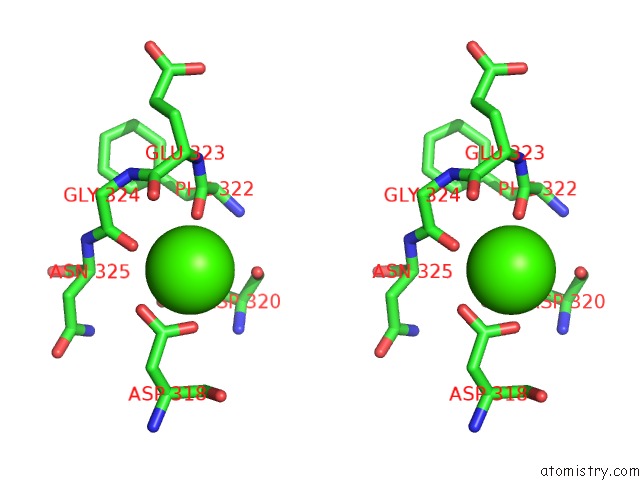
Calcium binding site 3 out of 4 in 1n86
Go back to
Calcium binding site 3 out
of 4 in the Crystal Structure of Human D-Dimer From Cross-Linked Fibrin Complexed with Gpr and Ghrpldk Peptide Ligands.
Mono view
Stereo pair view
Mono view
Stereo pair view
A full contact list of Calcium with other atoms in the Ca binding
site number 3 of Crystal Structure of Human D-Dimer From Cross-Linked Fibrin Complexed with Gpr and Ghrpldk Peptide Ligands. within 5.0Å range:
|
Calcium binding site 4 out of 4 in 1n86
Go back to
Calcium binding site 4 out
of 4 in the Crystal Structure of Human D-Dimer From Cross-Linked Fibrin Complexed with Gpr and Ghrpldk Peptide Ligands.
Mono view
Stereo pair view
Mono view
Stereo pair view
A full contact list of Calcium with other atoms in the Ca binding
site number 4 of Crystal Structure of Human D-Dimer From Cross-Linked Fibrin Complexed with Gpr and Ghrpldk Peptide Ligands. within 5.0Å range:
|
Reference:
Z.Yang,
L.Pandi,
R.F.Doolittle.
The Crystal Structure of Fragment Double-D From Cross-Linked Lamprey Fibrin Reveals Isopeptide Linkages Across An Unexpected D-D Interface. Biochemistry V. 41 15610 2002.
ISSN: ISSN 0006-2960
PubMed: 12501189
DOI: 10.1021/BI026666I
Page generated: Thu Jul 11 12:47:37 2024
ISSN: ISSN 0006-2960
PubMed: 12501189
DOI: 10.1021/BI026666I
Last articles
Zn in 9J0NZn in 9J0O
Zn in 9J0P
Zn in 9FJX
Zn in 9EKB
Zn in 9C0F
Zn in 9CAH
Zn in 9CH0
Zn in 9CH3
Zn in 9CH1