Calcium »
PDB 1n7s-1nm9 »
1n9e »
Calcium in PDB 1n9e: Crystal Structure of Pichia Pastoris Lysyl Oxidase Pplo
Enzymatic activity of Crystal Structure of Pichia Pastoris Lysyl Oxidase Pplo
All present enzymatic activity of Crystal Structure of Pichia Pastoris Lysyl Oxidase Pplo:
1.4.3.13;
1.4.3.13;
Protein crystallography data
The structure of Crystal Structure of Pichia Pastoris Lysyl Oxidase Pplo, PDB code: 1n9e
was solved by
J.M.Guss,
A.P.Duff,
with X-Ray Crystallography technique. A brief refinement statistics is given in the table below:
| Resolution Low / High (Å) | 24.60 / 1.65 |
| Space group | C 1 2 1 |
| Cell size a, b, c (Å), α, β, γ (°) | 248.442, 121.125, 151.841, 90.00, 124.64, 90.00 |
| R / Rfree (%) | 16.1 / 18.7 |
Other elements in 1n9e:
The structure of Crystal Structure of Pichia Pastoris Lysyl Oxidase Pplo also contains other interesting chemical elements:
| Copper | (Cu) | 4 atoms |
Calcium Binding Sites:
The binding sites of Calcium atom in the Crystal Structure of Pichia Pastoris Lysyl Oxidase Pplo
(pdb code 1n9e). This binding sites where shown within
5.0 Angstroms radius around Calcium atom.
In total 8 binding sites of Calcium where determined in the Crystal Structure of Pichia Pastoris Lysyl Oxidase Pplo, PDB code: 1n9e:
Jump to Calcium binding site number: 1; 2; 3; 4; 5; 6; 7; 8;
In total 8 binding sites of Calcium where determined in the Crystal Structure of Pichia Pastoris Lysyl Oxidase Pplo, PDB code: 1n9e:
Jump to Calcium binding site number: 1; 2; 3; 4; 5; 6; 7; 8;
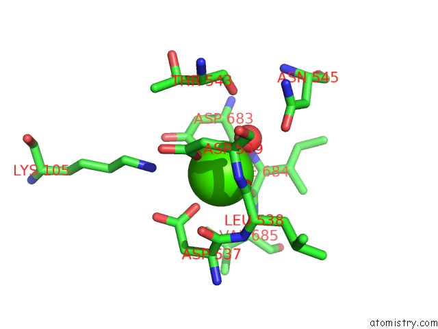
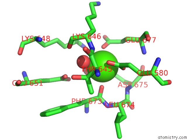
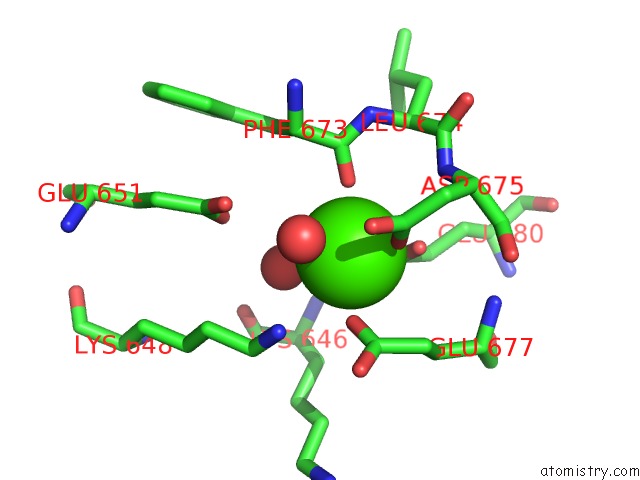
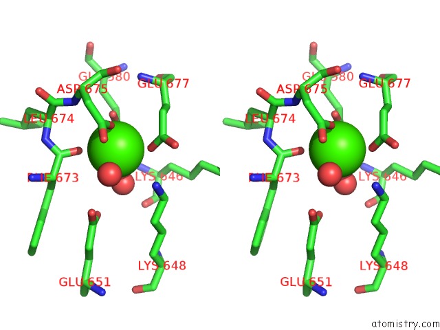
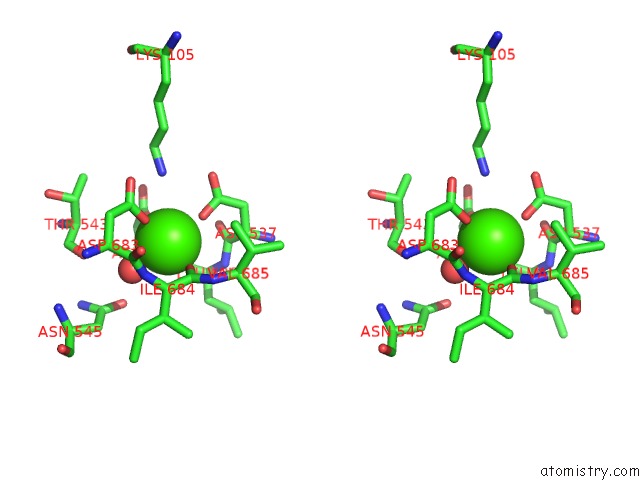
Calcium binding site 1 out of 8 in 1n9e
Go back to
Calcium binding site 1 out
of 8 in the Crystal Structure of Pichia Pastoris Lysyl Oxidase Pplo
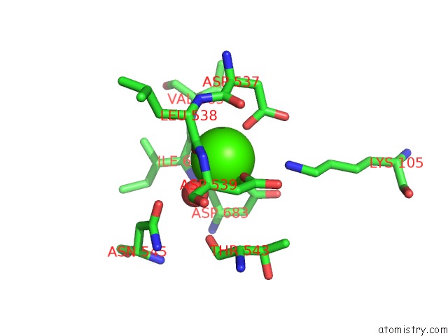
Mono view

Stereo pair view
Mono view
Stereo pair view
A full contact list of Calcium with other atoms in the Ca binding
site number 1 of Crystal Structure of Pichia Pastoris Lysyl Oxidase Pplo within 5.0Å range:
|
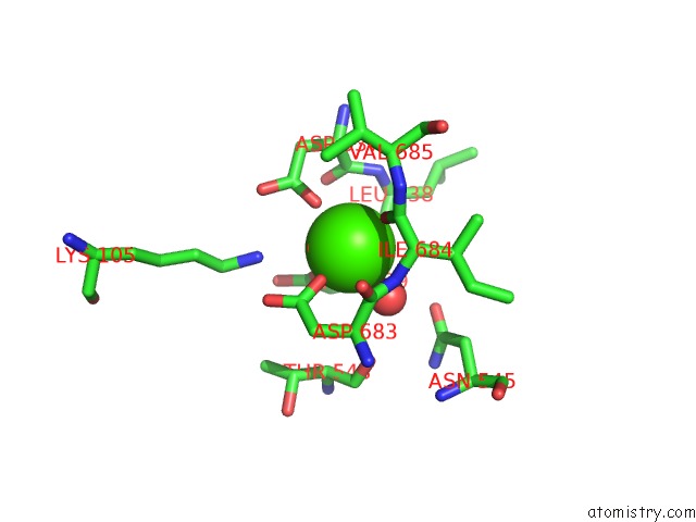
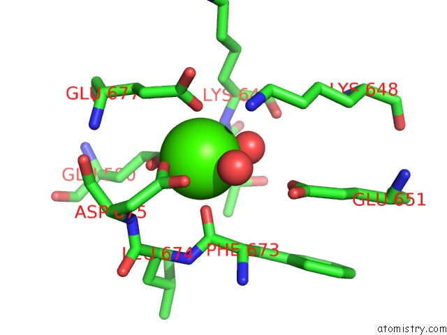
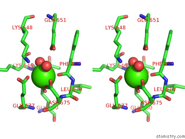
Calcium binding site 2 out of 8 in 1n9e
Go back to
Calcium binding site 2 out
of 8 in the Crystal Structure of Pichia Pastoris Lysyl Oxidase Pplo
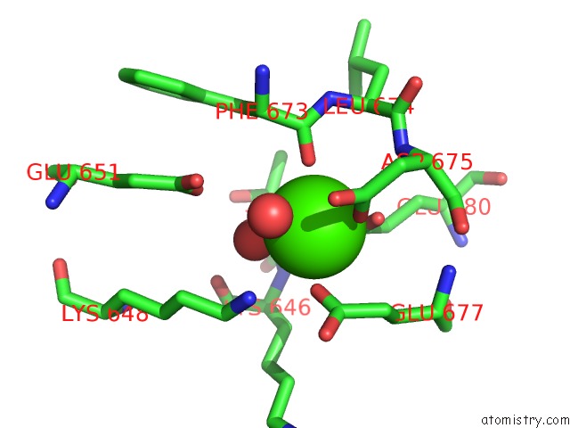
Mono view
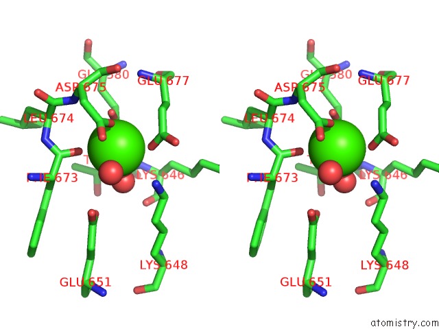
Stereo pair view
Mono view
Stereo pair view
A full contact list of Calcium with other atoms in the Ca binding
site number 2 of Crystal Structure of Pichia Pastoris Lysyl Oxidase Pplo within 5.0Å range:
|
Calcium binding site 3 out of 8 in 1n9e
Go back to
Calcium binding site 3 out
of 8 in the Crystal Structure of Pichia Pastoris Lysyl Oxidase Pplo
Mono view

Stereo pair view
Mono view
Stereo pair view
A full contact list of Calcium with other atoms in the Ca binding
site number 3 of Crystal Structure of Pichia Pastoris Lysyl Oxidase Pplo within 5.0Å range:
|
Calcium binding site 4 out of 8 in 1n9e
Go back to
Calcium binding site 4 out
of 8 in the Crystal Structure of Pichia Pastoris Lysyl Oxidase Pplo
Mono view

Stereo pair view
Mono view
Stereo pair view
A full contact list of Calcium with other atoms in the Ca binding
site number 4 of Crystal Structure of Pichia Pastoris Lysyl Oxidase Pplo within 5.0Å range:
|
Calcium binding site 5 out of 8 in 1n9e
Go back to
Calcium binding site 5 out
of 8 in the Crystal Structure of Pichia Pastoris Lysyl Oxidase Pplo
Mono view

Stereo pair view
Mono view
Stereo pair view
A full contact list of Calcium with other atoms in the Ca binding
site number 5 of Crystal Structure of Pichia Pastoris Lysyl Oxidase Pplo within 5.0Å range:
|
Calcium binding site 6 out of 8 in 1n9e
Go back to
Calcium binding site 6 out
of 8 in the Crystal Structure of Pichia Pastoris Lysyl Oxidase Pplo
Mono view
Stereo pair view
Mono view
Stereo pair view
A full contact list of Calcium with other atoms in the Ca binding
site number 6 of Crystal Structure of Pichia Pastoris Lysyl Oxidase Pplo within 5.0Å range:
|
Calcium binding site 7 out of 8 in 1n9e
Go back to
Calcium binding site 7 out
of 8 in the Crystal Structure of Pichia Pastoris Lysyl Oxidase Pplo

Mono view
Stereo pair view
Mono view
Stereo pair view
A full contact list of Calcium with other atoms in the Ca binding
site number 7 of Crystal Structure of Pichia Pastoris Lysyl Oxidase Pplo within 5.0Å range:
|
Calcium binding site 8 out of 8 in 1n9e
Go back to
Calcium binding site 8 out
of 8 in the Crystal Structure of Pichia Pastoris Lysyl Oxidase Pplo
Mono view
Stereo pair view
Mono view
Stereo pair view
A full contact list of Calcium with other atoms in the Ca binding
site number 8 of Crystal Structure of Pichia Pastoris Lysyl Oxidase Pplo within 5.0Å range:
|
Reference:
A.P.Duff,
A.E.Cohen,
P.J.Ellis,
J.A.Kuchar,
D.B.Langley,
E.M.Shepard,
D.M.Dooley,
H.C.Freeman,
J.M.Guss.
The Crystal Structure of Pichia Pastoris Lysyl Oxidase Biochemistry V. 42 15148 2003.
ISSN: ISSN 0006-2960
PubMed: 14690425
DOI: 10.1021/BI035338V
Page generated: Mon Jul 7 17:29:54 2025
ISSN: ISSN 0006-2960
PubMed: 14690425
DOI: 10.1021/BI035338V
Last articles
Cl in 5RU0Cl in 5RUH
Cl in 5RUO
Cl in 5RUA
Cl in 5RUF
Cl in 5RTX
Cl in 5RUD
Cl in 5RT6
Cl in 5RTH
Cl in 5RTI