Calcium »
PDB 1n7s-1nm9 »
1ncb »
Calcium in PDB 1ncb: Crystal Structures of Two Mutant Neuraminidase-Antibody Complexes with Amino Acid Substitutions in the Interface
Enzymatic activity of Crystal Structures of Two Mutant Neuraminidase-Antibody Complexes with Amino Acid Substitutions in the Interface
All present enzymatic activity of Crystal Structures of Two Mutant Neuraminidase-Antibody Complexes with Amino Acid Substitutions in the Interface:
3.2.1.18;
3.2.1.18;
Protein crystallography data
The structure of Crystal Structures of Two Mutant Neuraminidase-Antibody Complexes with Amino Acid Substitutions in the Interface, PDB code: 1ncb
was solved by
W.R.Tulip,
J.N.Varghese,
P.M.Colman,
with X-Ray Crystallography technique. A brief refinement statistics is given in the table below:
| Resolution Low / High (Å) | 8.00 / 2.50 |
| Space group | P 4 21 2 |
| Cell size a, b, c (Å), α, β, γ (°) | 167.000, 167.000, 124.000, 90.00, 90.00, 90.00 |
| R / Rfree (%) | 16.5 / n/a |
Calcium Binding Sites:
The binding sites of Calcium atom in the Crystal Structures of Two Mutant Neuraminidase-Antibody Complexes with Amino Acid Substitutions in the Interface
(pdb code 1ncb). This binding sites where shown within
5.0 Angstroms radius around Calcium atom.
In total only one binding site of Calcium was determined in the Crystal Structures of Two Mutant Neuraminidase-Antibody Complexes with Amino Acid Substitutions in the Interface, PDB code: 1ncb:
In total only one binding site of Calcium was determined in the Crystal Structures of Two Mutant Neuraminidase-Antibody Complexes with Amino Acid Substitutions in the Interface, PDB code: 1ncb:
Calcium binding site 1 out of 1 in 1ncb
Go back to
Calcium binding site 1 out
of 1 in the Crystal Structures of Two Mutant Neuraminidase-Antibody Complexes with Amino Acid Substitutions in the Interface
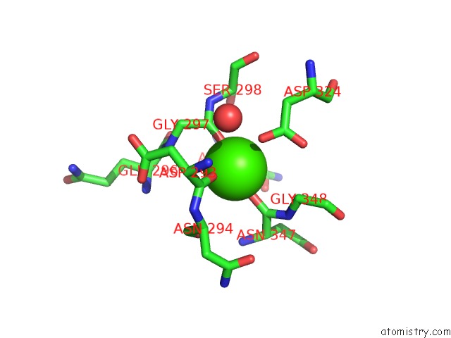
Mono view
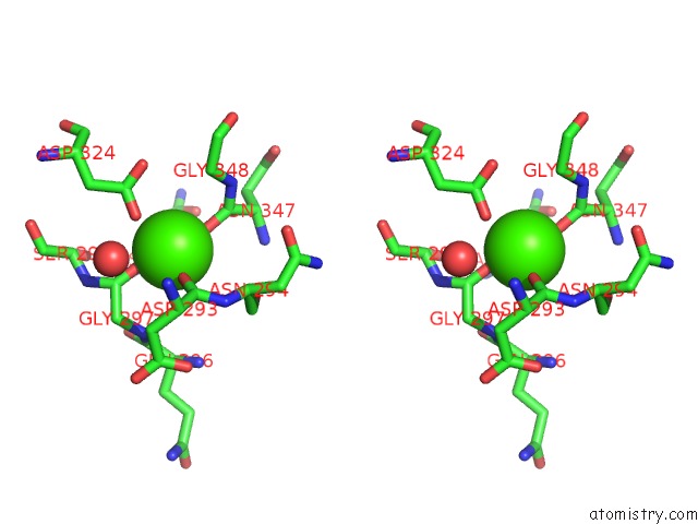
Stereo pair view
Mono view
Stereo pair view
A full contact list of Calcium with other atoms in the Ca binding
site number 1 of Crystal Structures of Two Mutant Neuraminidase-Antibody Complexes with Amino Acid Substitutions in the Interface within 5.0Å range:
|
Reference:
W.R.Tulip,
J.N.Varghese,
R.G.Webster,
W.G.Laver,
P.M.Colman.
Crystal Structures of Two Mutant Neuraminidase-Antibody Complexes with Amino Acid Substitutions in the Interface. J.Mol.Biol. V. 227 149 1992.
ISSN: ISSN 0022-2836
PubMed: 1522584
DOI: 10.1016/0022-2836(92)90688-G
Page generated: Thu Jul 11 12:48:20 2024
ISSN: ISSN 0022-2836
PubMed: 1522584
DOI: 10.1016/0022-2836(92)90688-G
Last articles
Zn in 9J0NZn in 9J0O
Zn in 9J0P
Zn in 9FJX
Zn in 9EKB
Zn in 9C0F
Zn in 9CAH
Zn in 9CH0
Zn in 9CH3
Zn in 9CH1