Calcium »
PDB 1n7s-1nm9 »
1nfw »
Calcium in PDB 1nfw: Crystal Structure of Human Coagulation Factor Xa Complexed with RPR209685
Enzymatic activity of Crystal Structure of Human Coagulation Factor Xa Complexed with RPR209685
All present enzymatic activity of Crystal Structure of Human Coagulation Factor Xa Complexed with RPR209685:
3.4.21.6;
3.4.21.6;
Protein crystallography data
The structure of Crystal Structure of Human Coagulation Factor Xa Complexed with RPR209685, PDB code: 1nfw
was solved by
S.Maignan,
J.P.Guilloteau,
with X-Ray Crystallography technique. A brief refinement statistics is given in the table below:
| Resolution Low / High (Å) | 20.00 / 2.10 |
| Space group | P 21 21 21 |
| Cell size a, b, c (Å), α, β, γ (°) | 55.950, 71.730, 78.960, 90.00, 90.00, 90.00 |
| R / Rfree (%) | 20 / n/a |
Other elements in 1nfw:
The structure of Crystal Structure of Human Coagulation Factor Xa Complexed with RPR209685 also contains other interesting chemical elements:
| Chlorine | (Cl) | 1 atom |
Calcium Binding Sites:
The binding sites of Calcium atom in the Crystal Structure of Human Coagulation Factor Xa Complexed with RPR209685
(pdb code 1nfw). This binding sites where shown within
5.0 Angstroms radius around Calcium atom.
In total only one binding site of Calcium was determined in the Crystal Structure of Human Coagulation Factor Xa Complexed with RPR209685, PDB code: 1nfw:
In total only one binding site of Calcium was determined in the Crystal Structure of Human Coagulation Factor Xa Complexed with RPR209685, PDB code: 1nfw:
Calcium binding site 1 out of 1 in 1nfw
Go back to
Calcium binding site 1 out
of 1 in the Crystal Structure of Human Coagulation Factor Xa Complexed with RPR209685
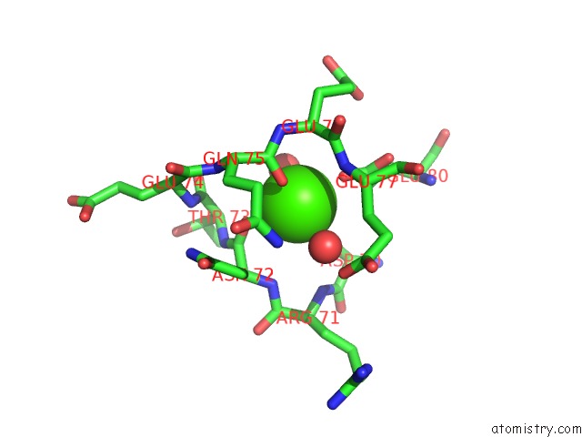
Mono view
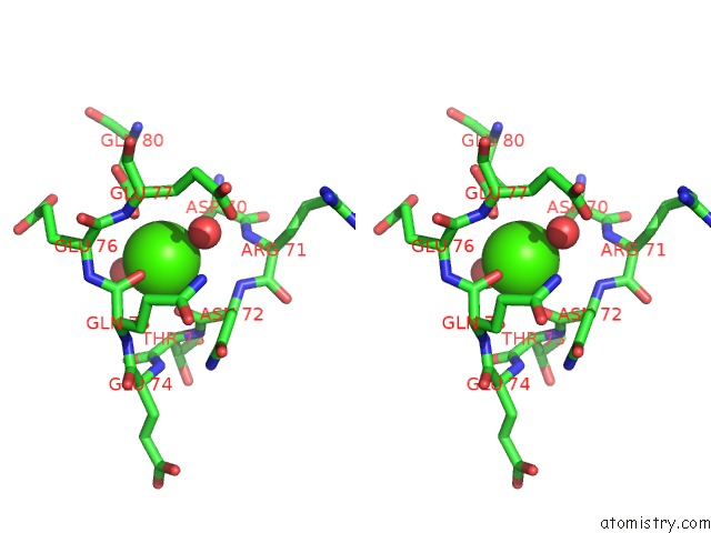
Stereo pair view
Mono view
Stereo pair view
A full contact list of Calcium with other atoms in the Ca binding
site number 1 of Crystal Structure of Human Coagulation Factor Xa Complexed with RPR209685 within 5.0Å range:
|
Reference:
S.Maignan,
J.P.Guilloteau,
Y.M.Choi-Sledeski,
M.R.Becker,
W.R.Ewing,
H.W.Pauls,
A.P.Spada,
V.Mikol.
Molecular Structures of Human Factor Xa Complexed with Ketopiperazine Inhibitors: Preference For A Neutral Group in the S1 Pocket. J.Med.Chem. V. 46 685 2003.
ISSN: ISSN 0022-2623
PubMed: 12593649
DOI: 10.1021/JM0203837
Page generated: Thu Jul 11 12:50:46 2024
ISSN: ISSN 0022-2623
PubMed: 12593649
DOI: 10.1021/JM0203837
Last articles
Zn in 9J0NZn in 9J0O
Zn in 9J0P
Zn in 9FJX
Zn in 9EKB
Zn in 9C0F
Zn in 9CAH
Zn in 9CH0
Zn in 9CH3
Zn in 9CH1