Calcium »
PDB 1n7s-1nm9 »
1nm1 »
Calcium in PDB 1nm1: Crystal Structure of D. Dicsoideum Actin Complexed with Gelsolin Segment 1 and Mg Atp at 1.8 A Resolution
Protein crystallography data
The structure of Crystal Structure of D. Dicsoideum Actin Complexed with Gelsolin Segment 1 and Mg Atp at 1.8 A Resolution, PDB code: 1nm1
was solved by
S.M.Vorobiev,
S.Welti,
J.Condeelis,
S.C.Almo,
with X-Ray Crystallography technique. A brief refinement statistics is given in the table below:
| Resolution Low / High (Å) | 20.00 / 1.80 |
| Space group | C 1 2 1 |
| Cell size a, b, c (Å), α, β, γ (°) | 178.539, 69.089, 56.552, 90.00, 104.34, 90.00 |
| R / Rfree (%) | 19.7 / 23.2 |
Other elements in 1nm1:
The structure of Crystal Structure of D. Dicsoideum Actin Complexed with Gelsolin Segment 1 and Mg Atp at 1.8 A Resolution also contains other interesting chemical elements:
| Magnesium | (Mg) | 1 atom |
Calcium Binding Sites:
The binding sites of Calcium atom in the Crystal Structure of D. Dicsoideum Actin Complexed with Gelsolin Segment 1 and Mg Atp at 1.8 A Resolution
(pdb code 1nm1). This binding sites where shown within
5.0 Angstroms radius around Calcium atom.
In total only one binding site of Calcium was determined in the Crystal Structure of D. Dicsoideum Actin Complexed with Gelsolin Segment 1 and Mg Atp at 1.8 A Resolution, PDB code: 1nm1:
In total only one binding site of Calcium was determined in the Crystal Structure of D. Dicsoideum Actin Complexed with Gelsolin Segment 1 and Mg Atp at 1.8 A Resolution, PDB code: 1nm1:
Calcium binding site 1 out of 1 in 1nm1
Go back to
Calcium binding site 1 out
of 1 in the Crystal Structure of D. Dicsoideum Actin Complexed with Gelsolin Segment 1 and Mg Atp at 1.8 A Resolution
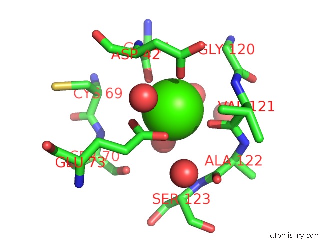
Mono view
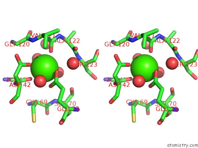
Stereo pair view
Mono view
Stereo pair view
A full contact list of Calcium with other atoms in the Ca binding
site number 1 of Crystal Structure of D. Dicsoideum Actin Complexed with Gelsolin Segment 1 and Mg Atp at 1.8 A Resolution within 5.0Å range:
|
Reference:
S.M.Vorobiev,
B.Strokopytov,
D.G.Drubin,
C.Frieden,
S.Ono,
J.Condeelis,
P.A.Rubenstein,
S.C.Almo.
The Structure of Non-Vertebrate Actin: Implications For the Atp Hydrolytic Mechanism Proc.Natl.Acad.Sci.Usa V. 100 5760 2003.
ISSN: ISSN 0027-8424
PubMed: 12732734
DOI: 10.1073/PNAS.0832273100
Page generated: Thu Jul 11 12:55:25 2024
ISSN: ISSN 0027-8424
PubMed: 12732734
DOI: 10.1073/PNAS.0832273100
Last articles
Zn in 9J0NZn in 9J0O
Zn in 9J0P
Zn in 9FJX
Zn in 9EKB
Zn in 9C0F
Zn in 9CAH
Zn in 9CH0
Zn in 9CH3
Zn in 9CH1