Calcium »
PDB 1nmb-1nxc »
1nps »
Calcium in PDB 1nps: Crystal Structure of N-Terminal Domain of Protein S
Protein crystallography data
The structure of Crystal Structure of N-Terminal Domain of Protein S, PDB code: 1nps
was solved by
M.Wenk,
R.Baumgartner,
E.M.Mayer,
R.Huber,
T.A.Holak,
R.Jaenicke,
with X-Ray Crystallography technique. A brief refinement statistics is given in the table below:
| Resolution Low / High (Å) | 20.00 / 1.80 |
| Space group | P 1 21 1 |
| Cell size a, b, c (Å), α, β, γ (°) | 28.360, 37.940, 37.260, 90.00, 105.93, 90.00 |
| R / Rfree (%) | 20 / 23.4 |
Calcium Binding Sites:
The binding sites of Calcium atom in the Crystal Structure of N-Terminal Domain of Protein S
(pdb code 1nps). This binding sites where shown within
5.0 Angstroms radius around Calcium atom.
In total 2 binding sites of Calcium where determined in the Crystal Structure of N-Terminal Domain of Protein S, PDB code: 1nps:
Jump to Calcium binding site number: 1; 2;
In total 2 binding sites of Calcium where determined in the Crystal Structure of N-Terminal Domain of Protein S, PDB code: 1nps:
Jump to Calcium binding site number: 1; 2;
Calcium binding site 1 out of 2 in 1nps
Go back to
Calcium binding site 1 out
of 2 in the Crystal Structure of N-Terminal Domain of Protein S
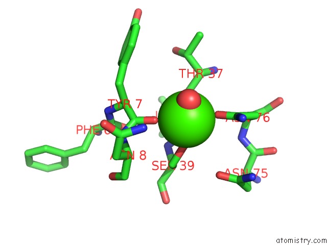
Mono view
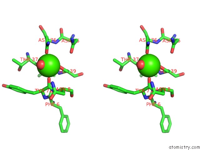
Stereo pair view
Mono view
Stereo pair view
A full contact list of Calcium with other atoms in the Ca binding
site number 1 of Crystal Structure of N-Terminal Domain of Protein S within 5.0Å range:
|
Calcium binding site 2 out of 2 in 1nps
Go back to
Calcium binding site 2 out
of 2 in the Crystal Structure of N-Terminal Domain of Protein S
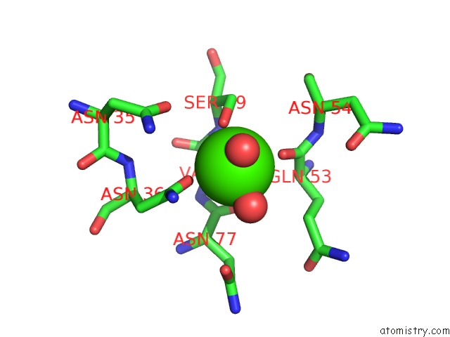
Mono view
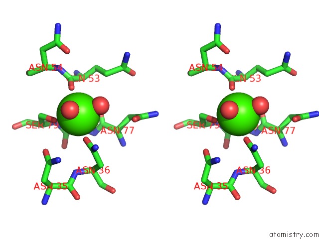
Stereo pair view
Mono view
Stereo pair view
A full contact list of Calcium with other atoms in the Ca binding
site number 2 of Crystal Structure of N-Terminal Domain of Protein S within 5.0Å range:
|
Reference:
M.Wenk,
R.Baumgartner,
T.A.Holak,
R.Huber,
R.Jaenicke,
E.M.Mayr.
The Domains of Protein S From Myxococcus Xanthus: Structure, Stability and Interactions. J.Mol.Biol. V. 286 1533 1999.
ISSN: ISSN 0022-2836
PubMed: 10064714
DOI: 10.1006/JMBI.1999.2582
Page generated: Thu Jul 11 12:58:27 2024
ISSN: ISSN 0022-2836
PubMed: 10064714
DOI: 10.1006/JMBI.1999.2582
Last articles
Zn in 9J0NZn in 9J0O
Zn in 9J0P
Zn in 9FJX
Zn in 9EKB
Zn in 9C0F
Zn in 9CAH
Zn in 9CH0
Zn in 9CH3
Zn in 9CH1