Calcium »
PDB 1nmb-1nxc »
1nt0 »
Calcium in PDB 1nt0: Crystal Structure of the CUB1-Egf-CUB2 Region of MASP2
Protein crystallography data
The structure of Crystal Structure of the CUB1-Egf-CUB2 Region of MASP2, PDB code: 1nt0
was solved by
H.Feinberg,
J.C.M.Uitdehaag,
J.M.Davies,
R.Wallis,
K.Drickamer,
W.I.Weis,
with X-Ray Crystallography technique. A brief refinement statistics is given in the table below:
| Resolution Low / High (Å) | 39.56 / 2.70 |
| Space group | P 1 21 1 |
| Cell size a, b, c (Å), α, β, γ (°) | 70.411, 103.901, 70.484, 90.00, 119.93, 90.00 |
| R / Rfree (%) | 24.8 / 28.3 |
Calcium Binding Sites:
The binding sites of Calcium atom in the Crystal Structure of the CUB1-Egf-CUB2 Region of MASP2
(pdb code 1nt0). This binding sites where shown within
5.0 Angstroms radius around Calcium atom.
In total 2 binding sites of Calcium where determined in the Crystal Structure of the CUB1-Egf-CUB2 Region of MASP2, PDB code: 1nt0:
Jump to Calcium binding site number: 1; 2;
In total 2 binding sites of Calcium where determined in the Crystal Structure of the CUB1-Egf-CUB2 Region of MASP2, PDB code: 1nt0:
Jump to Calcium binding site number: 1; 2;
Calcium binding site 1 out of 2 in 1nt0
Go back to
Calcium binding site 1 out
of 2 in the Crystal Structure of the CUB1-Egf-CUB2 Region of MASP2
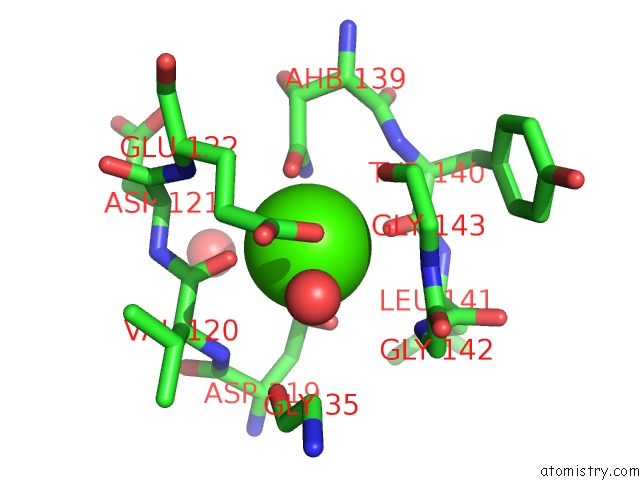
Mono view
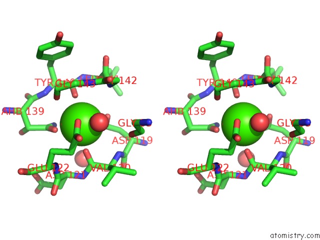
Stereo pair view
Mono view
Stereo pair view
A full contact list of Calcium with other atoms in the Ca binding
site number 1 of Crystal Structure of the CUB1-Egf-CUB2 Region of MASP2 within 5.0Å range:
|
Calcium binding site 2 out of 2 in 1nt0
Go back to
Calcium binding site 2 out
of 2 in the Crystal Structure of the CUB1-Egf-CUB2 Region of MASP2
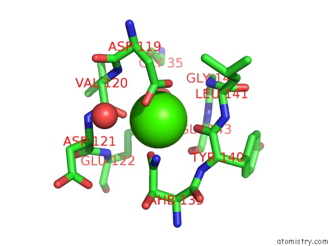
Mono view
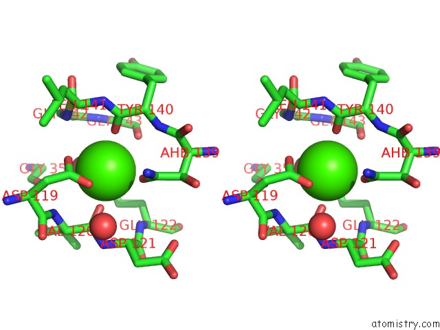
Stereo pair view
Mono view
Stereo pair view
A full contact list of Calcium with other atoms in the Ca binding
site number 2 of Crystal Structure of the CUB1-Egf-CUB2 Region of MASP2 within 5.0Å range:
|
Reference:
H.Feinberg,
J.C.M.Uitdehaag,
J.M.Davies,
R.Wallis,
K.Drickamer,
W.I.Weis.
Crystal Structure of the CUB1-Egf-CUB2 Region of Mannose-Binding Protein Associated Serine Protease-2 Embo J. V. 22 2348 2003.
ISSN: ISSN 0261-4189
PubMed: 12743029
DOI: 10.1093/EMBOJ/CDG236
Page generated: Thu Jul 11 13:01:25 2024
ISSN: ISSN 0261-4189
PubMed: 12743029
DOI: 10.1093/EMBOJ/CDG236
Last articles
Zn in 9J0NZn in 9J0O
Zn in 9J0P
Zn in 9FJX
Zn in 9EKB
Zn in 9C0F
Zn in 9CAH
Zn in 9CH0
Zn in 9CH3
Zn in 9CH1