Calcium »
PDB 1nmb-1nxc »
1nxc »
Calcium in PDB 1nxc: Structure of Mouse Golgi Alpha-1,2-Mannosidase Ia Reveals the Molecular Basis For Substrate Specificity Among Class I Enzymes (Family 47 Glycosidases)
Enzymatic activity of Structure of Mouse Golgi Alpha-1,2-Mannosidase Ia Reveals the Molecular Basis For Substrate Specificity Among Class I Enzymes (Family 47 Glycosidases)
All present enzymatic activity of Structure of Mouse Golgi Alpha-1,2-Mannosidase Ia Reveals the Molecular Basis For Substrate Specificity Among Class I Enzymes (Family 47 Glycosidases):
3.2.1.113;
3.2.1.113;
Protein crystallography data
The structure of Structure of Mouse Golgi Alpha-1,2-Mannosidase Ia Reveals the Molecular Basis For Substrate Specificity Among Class I Enzymes (Family 47 Glycosidases), PDB code: 1nxc
was solved by
W.Tempel,
Z.-J.Liu,
K.Karaveg,
J.Rose,
K.W.Moremen,
B.-C.Wang,
Southeastcollaboratory For Structural Genomics (Secsg),
with X-Ray Crystallography technique. A brief refinement statistics is given in the table below:
| Resolution Low / High (Å) | 62.50 / 1.51 |
| Space group | P 21 21 21 |
| Cell size a, b, c (Å), α, β, γ (°) | 55.288, 72.164, 129.571, 90.00, 90.00, 90.00 |
| R / Rfree (%) | 17.1 / 19 |
Calcium Binding Sites:
The binding sites of Calcium atom in the Structure of Mouse Golgi Alpha-1,2-Mannosidase Ia Reveals the Molecular Basis For Substrate Specificity Among Class I Enzymes (Family 47 Glycosidases)
(pdb code 1nxc). This binding sites where shown within
5.0 Angstroms radius around Calcium atom.
In total only one binding site of Calcium was determined in the Structure of Mouse Golgi Alpha-1,2-Mannosidase Ia Reveals the Molecular Basis For Substrate Specificity Among Class I Enzymes (Family 47 Glycosidases), PDB code: 1nxc:
In total only one binding site of Calcium was determined in the Structure of Mouse Golgi Alpha-1,2-Mannosidase Ia Reveals the Molecular Basis For Substrate Specificity Among Class I Enzymes (Family 47 Glycosidases), PDB code: 1nxc:
Calcium binding site 1 out of 1 in 1nxc
Go back to
Calcium binding site 1 out
of 1 in the Structure of Mouse Golgi Alpha-1,2-Mannosidase Ia Reveals the Molecular Basis For Substrate Specificity Among Class I Enzymes (Family 47 Glycosidases)
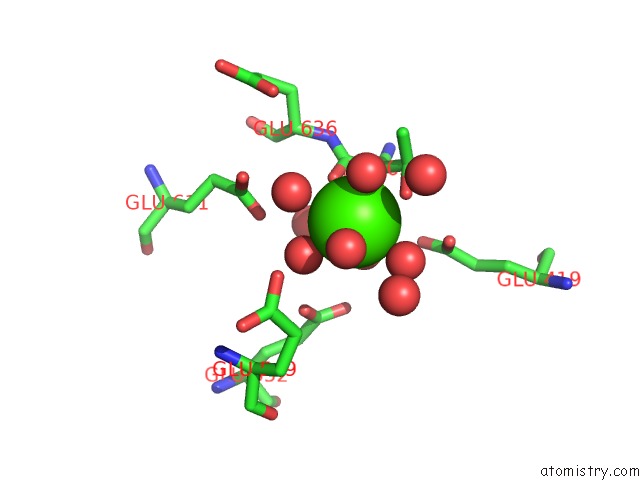
Mono view
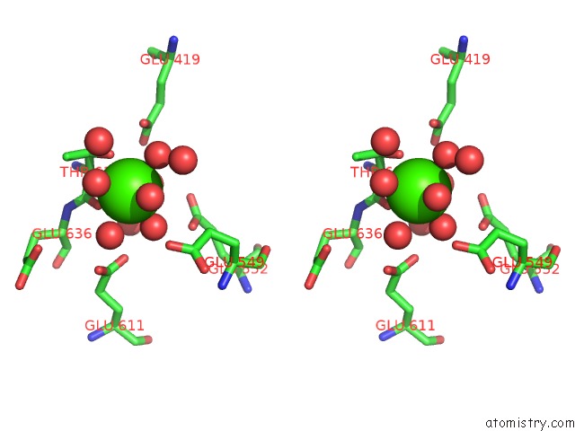
Stereo pair view
Mono view
Stereo pair view
A full contact list of Calcium with other atoms in the Ca binding
site number 1 of Structure of Mouse Golgi Alpha-1,2-Mannosidase Ia Reveals the Molecular Basis For Substrate Specificity Among Class I Enzymes (Family 47 Glycosidases) within 5.0Å range:
|
Reference:
W.Tempel,
K.Karaveg,
Z.-J.Liu,
J.Rose,
B.-C.Wang,
K.W.Moremen.
Structure of Mouse Golgi Alpha-Mannosidase Ia Reveals the Molecular Basis For Substrate Specificity Among Class 1 (Family 47 Glycosylhydrolase) ALPHA1,2-Mannosidases J.Biol.Chem. V. 279 29774 2004.
ISSN: ISSN 0021-9258
PubMed: 15102839
DOI: 10.1074/JBC.M403065200
Page generated: Thu Jul 11 13:05:46 2024
ISSN: ISSN 0021-9258
PubMed: 15102839
DOI: 10.1074/JBC.M403065200
Last articles
Zn in 9J0NZn in 9J0O
Zn in 9J0P
Zn in 9FJX
Zn in 9EKB
Zn in 9C0F
Zn in 9CAH
Zn in 9CH0
Zn in 9CH3
Zn in 9CH1