Calcium »
PDB 1o3d-1odb »
1o4y »
Calcium in PDB 1o4y: The Three-Dimensional Structure of Beta-Agarase A From Zobellia Galactanivorans
Enzymatic activity of The Three-Dimensional Structure of Beta-Agarase A From Zobellia Galactanivorans
All present enzymatic activity of The Three-Dimensional Structure of Beta-Agarase A From Zobellia Galactanivorans:
3.2.1.81;
3.2.1.81;
Protein crystallography data
The structure of The Three-Dimensional Structure of Beta-Agarase A From Zobellia Galactanivorans, PDB code: 1o4y
was solved by
J.Allouch,
M.Jam,
W.Helbert,
T.Barbeyron,
B.Kloareg,
B.Henrissat,
M.Czjzek,
with X-Ray Crystallography technique. A brief refinement statistics is given in the table below:
| Resolution Low / High (Å) | 34.00 / 1.48 |
| Space group | P 21 21 21 |
| Cell size a, b, c (Å), α, β, γ (°) | 50.314, 57.487, 89.054, 90.00, 90.00, 90.00 |
| R / Rfree (%) | 14.8 / 17.6 |
Other elements in 1o4y:
The structure of The Three-Dimensional Structure of Beta-Agarase A From Zobellia Galactanivorans also contains other interesting chemical elements:
| Sodium | (Na) | 1 atom |
Calcium Binding Sites:
The binding sites of Calcium atom in the The Three-Dimensional Structure of Beta-Agarase A From Zobellia Galactanivorans
(pdb code 1o4y). This binding sites where shown within
5.0 Angstroms radius around Calcium atom.
In total only one binding site of Calcium was determined in the The Three-Dimensional Structure of Beta-Agarase A From Zobellia Galactanivorans, PDB code: 1o4y:
In total only one binding site of Calcium was determined in the The Three-Dimensional Structure of Beta-Agarase A From Zobellia Galactanivorans, PDB code: 1o4y:
Calcium binding site 1 out of 1 in 1o4y
Go back to
Calcium binding site 1 out
of 1 in the The Three-Dimensional Structure of Beta-Agarase A From Zobellia Galactanivorans
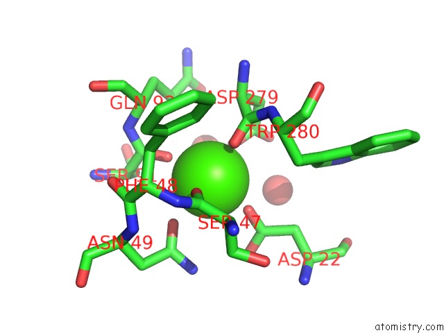
Mono view
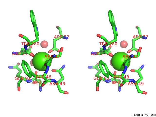
Stereo pair view
Mono view
Stereo pair view
A full contact list of Calcium with other atoms in the Ca binding
site number 1 of The Three-Dimensional Structure of Beta-Agarase A From Zobellia Galactanivorans within 5.0Å range:
|
Reference:
J.Allouch,
M.Jam,
W.Helbert,
T.Barbeyron,
B.Kloareg,
B.Henrissat,
M.Czjzek.
The Three-Dimensional Structures of Two {Beta}-Agarases. J.Biol.Chem. V. 278 47171 2003.
ISSN: ISSN 0021-9258
PubMed: 12970344
DOI: 10.1074/JBC.M308313200
Page generated: Mon Jul 7 17:50:38 2025
ISSN: ISSN 0021-9258
PubMed: 12970344
DOI: 10.1074/JBC.M308313200
Last articles
Ca in 7F9BCa in 7F98
Ca in 7F99
Ca in 7F8L
Ca in 7F7R
Ca in 7F5F
Ca in 7F5B
Ca in 7F58
Ca in 7F55
Ca in 7F54