Calcium »
PDB 1o3d-1odb »
1o8s »
Calcium in PDB 1o8s: Structure of CSCBM6-3 From Clostridium Stercorarium in Complex with Cellobiose
Enzymatic activity of Structure of CSCBM6-3 From Clostridium Stercorarium in Complex with Cellobiose
All present enzymatic activity of Structure of CSCBM6-3 From Clostridium Stercorarium in Complex with Cellobiose:
3.2.1.8;
3.2.1.8;
Protein crystallography data
The structure of Structure of CSCBM6-3 From Clostridium Stercorarium in Complex with Cellobiose, PDB code: 1o8s
was solved by
A.B.Boraston,
V.Notenboom,
R.A.J.Warren,
D.G.Kilbrun,
D.R.Rose,
G.J.Davies,
with X-Ray Crystallography technique. A brief refinement statistics is given in the table below:
| Resolution Low / High (Å) | 40.49 / 1.15 |
| Space group | P 21 21 21 |
| Cell size a, b, c (Å), α, β, γ (°) | 36.090, 52.069, 64.743, 90.00, 90.00, 90.00 |
| R / Rfree (%) | 12.3 / 14.9 |
Calcium Binding Sites:
The binding sites of Calcium atom in the Structure of CSCBM6-3 From Clostridium Stercorarium in Complex with Cellobiose
(pdb code 1o8s). This binding sites where shown within
5.0 Angstroms radius around Calcium atom.
In total only one binding site of Calcium was determined in the Structure of CSCBM6-3 From Clostridium Stercorarium in Complex with Cellobiose, PDB code: 1o8s:
In total only one binding site of Calcium was determined in the Structure of CSCBM6-3 From Clostridium Stercorarium in Complex with Cellobiose, PDB code: 1o8s:
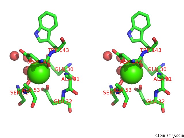
Calcium binding site 1 out of 1 in 1o8s
Go back to
Calcium binding site 1 out
of 1 in the Structure of CSCBM6-3 From Clostridium Stercorarium in Complex with Cellobiose

Mono view
Stereo pair view
Mono view
Stereo pair view
A full contact list of Calcium with other atoms in the Ca binding
site number 1 of Structure of CSCBM6-3 From Clostridium Stercorarium in Complex with Cellobiose within 5.0Å range:
|
Reference:
A.B.Boraston,
V.Notenboom,
R.A.J.Warren,
D.G.Kilburn,
D.R.Rose,
G.J.Davies.
Structure and Ligand Binding of Carbohydrate-Binding Module CSCBM6-3 Reveals Similarities with Fucose-Specific Lectins and Galactose-Binding Domains J.Mol.Biol. V. 327 659 2003.
ISSN: ISSN 0022-2836
PubMed: 12634060
DOI: 10.1016/S0022-2836(03)00152-9
Page generated: Mon Jul 7 17:51:45 2025
ISSN: ISSN 0022-2836
PubMed: 12634060
DOI: 10.1016/S0022-2836(03)00152-9
Last articles
Ca in 1Q3ACa in 1Q39
Ca in 1Q4N
Ca in 1Q1P
Ca in 1PZ8
Ca in 1Q2E
Ca in 1Q23
Ca in 1PZY
Ca in 1PZ7
Ca in 1PYT