Calcium »
PDB 1of3-1oux »
1okg »
Calcium in PDB 1okg: 3-Mercaptopyruvate Sulfurtransferase From Leishmania Major
Enzymatic activity of 3-Mercaptopyruvate Sulfurtransferase From Leishmania Major
All present enzymatic activity of 3-Mercaptopyruvate Sulfurtransferase From Leishmania Major:
2.8.1.2;
2.8.1.2;
Protein crystallography data
The structure of 3-Mercaptopyruvate Sulfurtransferase From Leishmania Major, PDB code: 1okg
was solved by
M.S.Alphey,
W.N.Hunter,
with X-Ray Crystallography technique. A brief refinement statistics is given in the table below:
| Resolution Low / High (Å) | 76.70 / 2.10 |
| Space group | P 42 21 2 |
| Cell size a, b, c (Å), α, β, γ (°) | 109.574, 109.574, 67.300, 90.00, 90.00, 90.00 |
| R / Rfree (%) | 20.8 / 28.7 |
Other elements in 1okg:
The structure of 3-Mercaptopyruvate Sulfurtransferase From Leishmania Major also contains other interesting chemical elements:
| Arsenic | (As) | 2 atoms |
Calcium Binding Sites:
The binding sites of Calcium atom in the 3-Mercaptopyruvate Sulfurtransferase From Leishmania Major
(pdb code 1okg). This binding sites where shown within
5.0 Angstroms radius around Calcium atom.
In total only one binding site of Calcium was determined in the 3-Mercaptopyruvate Sulfurtransferase From Leishmania Major, PDB code: 1okg:
In total only one binding site of Calcium was determined in the 3-Mercaptopyruvate Sulfurtransferase From Leishmania Major, PDB code: 1okg:
Calcium binding site 1 out of 1 in 1okg
Go back to
Calcium binding site 1 out
of 1 in the 3-Mercaptopyruvate Sulfurtransferase From Leishmania Major
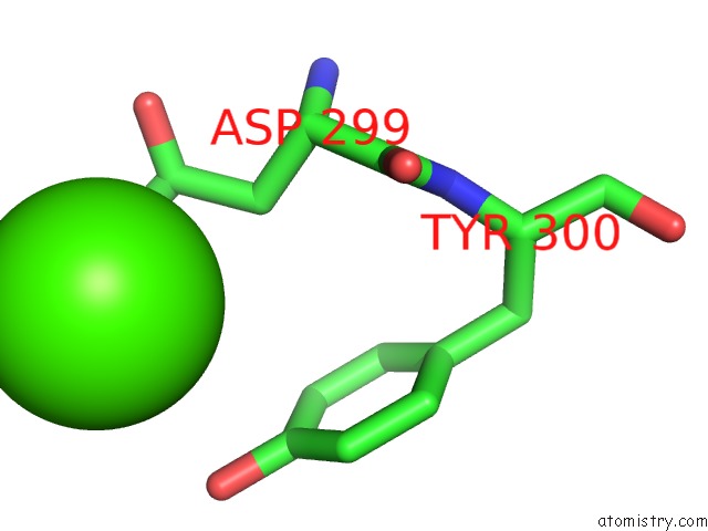
Mono view
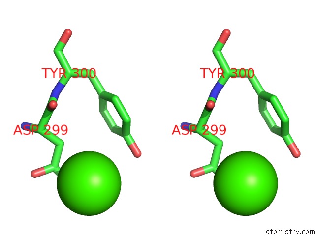
Stereo pair view
Mono view
Stereo pair view
A full contact list of Calcium with other atoms in the Ca binding
site number 1 of 3-Mercaptopyruvate Sulfurtransferase From Leishmania Major within 5.0Å range:
|
Reference:
M.S.Alphey,
R.A.M.Williams,
J.C.Mottram,
G.H.Coombs,
W.N.Hunter.
The Crystal Structure of Leishmania Major 3-Mercaptopyruvate Sulfurtransferase: A Three-Domain Architecture with A Serine Protease-Like Triad at the Active Site J.Biol.Chem. V. 278 48219 2003.
ISSN: ISSN 0021-9258
PubMed: 12952945
DOI: 10.1074/JBC.M307187200
Page generated: Thu Jul 11 13:22:30 2024
ISSN: ISSN 0021-9258
PubMed: 12952945
DOI: 10.1074/JBC.M307187200
Last articles
Zn in 9JYWZn in 9IR4
Zn in 9IR3
Zn in 9GMX
Zn in 9GMW
Zn in 9JEJ
Zn in 9ERF
Zn in 9ERE
Zn in 9EGV
Zn in 9EGW