Calcium »
PDB 1of3-1oux »
1os1 »
Calcium in PDB 1os1: Structure of Phosphoenolpyruvate Carboxykinase Complexed with Atp,Mg, Ca and Pyruvate.
Enzymatic activity of Structure of Phosphoenolpyruvate Carboxykinase Complexed with Atp,Mg, Ca and Pyruvate.
All present enzymatic activity of Structure of Phosphoenolpyruvate Carboxykinase Complexed with Atp,Mg, Ca and Pyruvate.:
4.1.1.49;
4.1.1.49;
Protein crystallography data
The structure of Structure of Phosphoenolpyruvate Carboxykinase Complexed with Atp,Mg, Ca and Pyruvate., PDB code: 1os1
was solved by
A.Sudom,
R.Walters,
L.Pastushok,
D.Goldie,
L.Prasad,
L.T.Delbaere,
H.Goldie,
with X-Ray Crystallography technique. A brief refinement statistics is given in the table below:
| Resolution Low / High (Å) | 10.00 / 1.80 |
| Space group | C 1 2 1 |
| Cell size a, b, c (Å), α, β, γ (°) | 126.200, 95.200, 46.800, 90.00, 95.20, 90.00 |
| R / Rfree (%) | 20 / 25 |
Other elements in 1os1:
The structure of Structure of Phosphoenolpyruvate Carboxykinase Complexed with Atp,Mg, Ca and Pyruvate. also contains other interesting chemical elements:
| Magnesium | (Mg) | 1 atom |
Calcium Binding Sites:
The binding sites of Calcium atom in the Structure of Phosphoenolpyruvate Carboxykinase Complexed with Atp,Mg, Ca and Pyruvate.
(pdb code 1os1). This binding sites where shown within
5.0 Angstroms radius around Calcium atom.
In total only one binding site of Calcium was determined in the Structure of Phosphoenolpyruvate Carboxykinase Complexed with Atp,Mg, Ca and Pyruvate., PDB code: 1os1:
In total only one binding site of Calcium was determined in the Structure of Phosphoenolpyruvate Carboxykinase Complexed with Atp,Mg, Ca and Pyruvate., PDB code: 1os1:
Calcium binding site 1 out of 1 in 1os1
Go back to
Calcium binding site 1 out
of 1 in the Structure of Phosphoenolpyruvate Carboxykinase Complexed with Atp,Mg, Ca and Pyruvate.
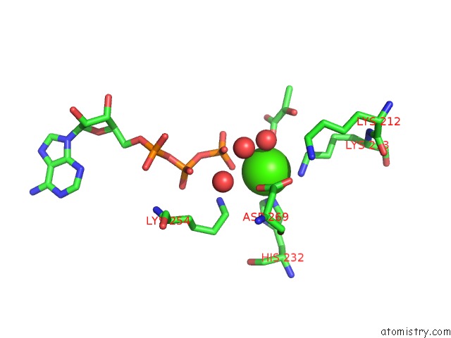
Mono view
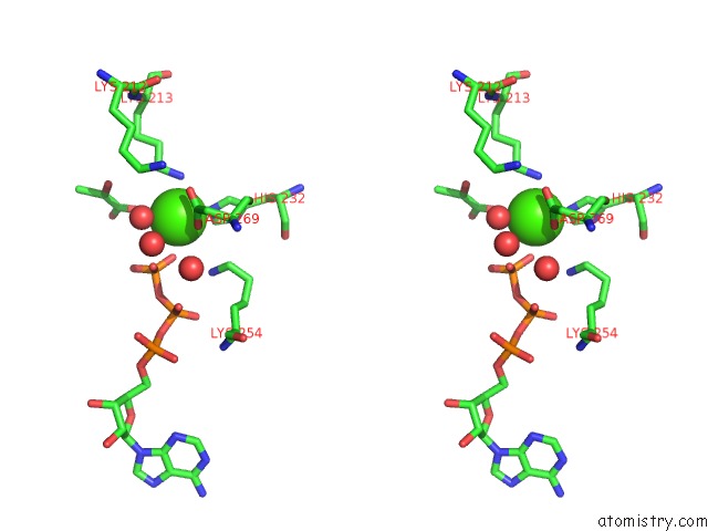
Stereo pair view
Mono view
Stereo pair view
A full contact list of Calcium with other atoms in the Ca binding
site number 1 of Structure of Phosphoenolpyruvate Carboxykinase Complexed with Atp,Mg, Ca and Pyruvate. within 5.0Å range:
|
Reference:
A.Sudom,
R.Walters,
L.Pastushok,
D.Goldie,
L.Prasad,
L.T.Delbaere,
H.Goldie.
Mechanisms of Activation of Phosphoenolpyruvate Carboxykinase From Escherichia Coli By CA2+ and of Desensitization By Trypsin. J.Bacteriol. V. 185 4233 2003.
ISSN: ISSN 0021-9193
PubMed: 12837799
DOI: 10.1128/JB.185.14.4233-4242.2003
Page generated: Thu Jul 11 13:28:37 2024
ISSN: ISSN 0021-9193
PubMed: 12837799
DOI: 10.1128/JB.185.14.4233-4242.2003
Last articles
Zn in 9J0NZn in 9J0O
Zn in 9J0P
Zn in 9FJX
Zn in 9EKB
Zn in 9C0F
Zn in 9CAH
Zn in 9CH0
Zn in 9CH3
Zn in 9CH1