Calcium »
PDB 1of3-1oux »
1ou9 »
Calcium in PDB 1ou9: Structure of Sspb, A Aaa+ Protease Delivery Protein
Protein crystallography data
The structure of Structure of Sspb, A Aaa+ Protease Delivery Protein, PDB code: 1ou9
was solved by
I.Levchenko,
R.A.Grant,
D.A.Wah,
R.T.Sauer,
T.A.Baker,
with X-Ray Crystallography technique. A brief refinement statistics is given in the table below:
| Resolution Low / High (Å) | 16.95 / 1.80 |
| Space group | C 2 2 21 |
| Cell size a, b, c (Å), α, β, γ (°) | 65.292, 137.839, 93.790, 90.00, 90.00, 90.00 |
| R / Rfree (%) | 21.7 / 25.3 |
Calcium Binding Sites:
The binding sites of Calcium atom in the Structure of Sspb, A Aaa+ Protease Delivery Protein
(pdb code 1ou9). This binding sites where shown within
5.0 Angstroms radius around Calcium atom.
In total 2 binding sites of Calcium where determined in the Structure of Sspb, A Aaa+ Protease Delivery Protein, PDB code: 1ou9:
Jump to Calcium binding site number: 1; 2;
In total 2 binding sites of Calcium where determined in the Structure of Sspb, A Aaa+ Protease Delivery Protein, PDB code: 1ou9:
Jump to Calcium binding site number: 1; 2;
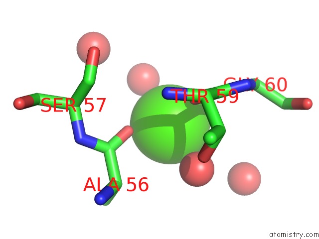
Calcium binding site 1 out of 2 in 1ou9
Go back to
Calcium binding site 1 out
of 2 in the Structure of Sspb, A Aaa+ Protease Delivery Protein
Mono view

Stereo pair view
Mono view
Stereo pair view
A full contact list of Calcium with other atoms in the Ca binding
site number 1 of Structure of Sspb, A Aaa+ Protease Delivery Protein within 5.0Å range:
|
Calcium binding site 2 out of 2 in 1ou9
Go back to
Calcium binding site 2 out
of 2 in the Structure of Sspb, A Aaa+ Protease Delivery Protein

Mono view

Stereo pair view
Mono view
Stereo pair view
A full contact list of Calcium with other atoms in the Ca binding
site number 2 of Structure of Sspb, A Aaa+ Protease Delivery Protein within 5.0Å range:
|
Reference:
I.Levchenko,
R.A.Grant,
D.A.Wah,
R.T.Sauer,
T.A.Baker.
Structure of A Delivery Protein For An Aaa+ Protease in Complex with A Peptide Degradation Tag Mol.Cell V. 12 365 2003.
ISSN: ISSN 1097-2765
PubMed: 14536076
DOI: 10.1016/J.MOLCEL.2003.08.014
Page generated: Thu Jul 11 13:31:19 2024
ISSN: ISSN 1097-2765
PubMed: 14536076
DOI: 10.1016/J.MOLCEL.2003.08.014
Last articles
Zn in 9J0NZn in 9J0O
Zn in 9J0P
Zn in 9FJX
Zn in 9EKB
Zn in 9C0F
Zn in 9CAH
Zn in 9CH0
Zn in 9CH3
Zn in 9CH1