Calcium »
PDB 1ova-1pif »
1pam »
Calcium in PDB 1pam: Cyclodextrin Glucanotransferase
Enzymatic activity of Cyclodextrin Glucanotransferase
All present enzymatic activity of Cyclodextrin Glucanotransferase:
2.4.1.19;
2.4.1.19;
Protein crystallography data
The structure of Cyclodextrin Glucanotransferase, PDB code: 1pam
was solved by
K.Harata,
K.Haga,
A.Nakamura,
M.Aoyagi,
K.Yamane,
with X-Ray Crystallography technique. A brief refinement statistics is given in the table below:
| Resolution Low / High (Å) | 10.00 / 1.80 |
| Space group | P 1 |
| Cell size a, b, c (Å), α, β, γ (°) | 64.930, 74.450, 79.120, 85.20, 105.00, 101.00 |
| R / Rfree (%) | 16.1 / 21.1 |
Calcium Binding Sites:
The binding sites of Calcium atom in the Cyclodextrin Glucanotransferase
(pdb code 1pam). This binding sites where shown within
5.0 Angstroms radius around Calcium atom.
In total 4 binding sites of Calcium where determined in the Cyclodextrin Glucanotransferase, PDB code: 1pam:
Jump to Calcium binding site number: 1; 2; 3; 4;
In total 4 binding sites of Calcium where determined in the Cyclodextrin Glucanotransferase, PDB code: 1pam:
Jump to Calcium binding site number: 1; 2; 3; 4;
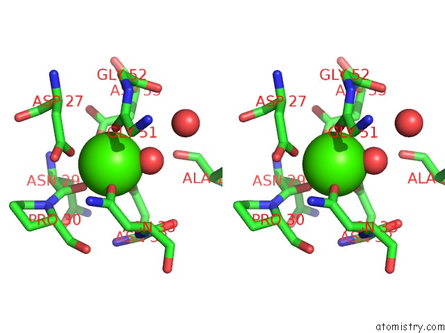
Calcium binding site 1 out of 4 in 1pam
Go back to
Calcium binding site 1 out
of 4 in the Cyclodextrin Glucanotransferase

Mono view
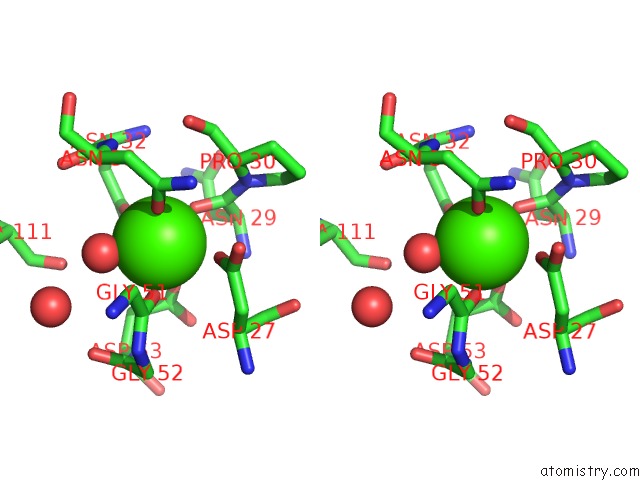
Stereo pair view
Mono view
Stereo pair view
A full contact list of Calcium with other atoms in the Ca binding
site number 1 of Cyclodextrin Glucanotransferase within 5.0Å range:
|
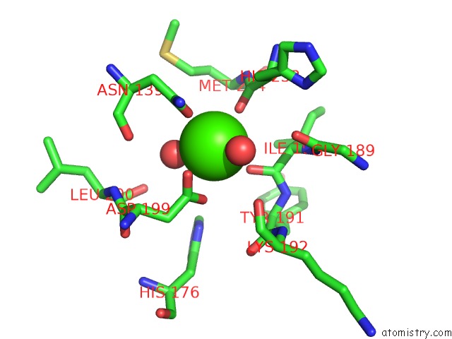
Calcium binding site 2 out of 4 in 1pam
Go back to
Calcium binding site 2 out
of 4 in the Cyclodextrin Glucanotransferase
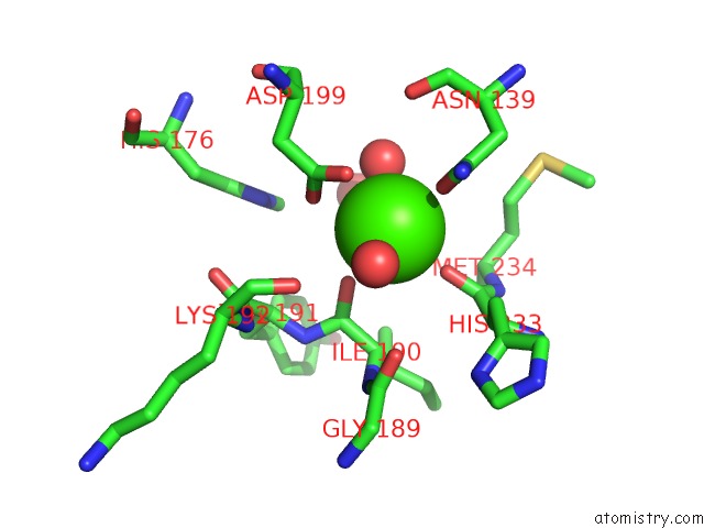
Mono view
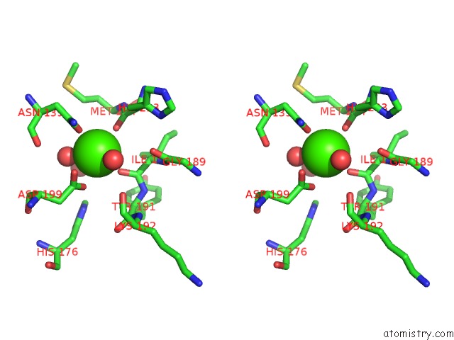
Stereo pair view
Mono view
Stereo pair view
A full contact list of Calcium with other atoms in the Ca binding
site number 2 of Cyclodextrin Glucanotransferase within 5.0Å range:
|
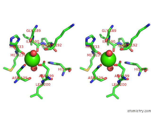
Calcium binding site 3 out of 4 in 1pam
Go back to
Calcium binding site 3 out
of 4 in the Cyclodextrin Glucanotransferase
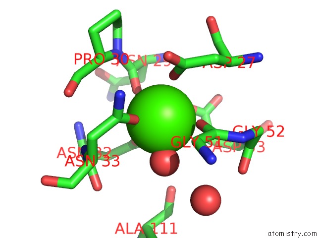
Mono view
Stereo pair view
Mono view
Stereo pair view
A full contact list of Calcium with other atoms in the Ca binding
site number 3 of Cyclodextrin Glucanotransferase within 5.0Å range:
|
Calcium binding site 4 out of 4 in 1pam
Go back to
Calcium binding site 4 out
of 4 in the Cyclodextrin Glucanotransferase
Mono view
Stereo pair view
Mono view
Stereo pair view
A full contact list of Calcium with other atoms in the Ca binding
site number 4 of Cyclodextrin Glucanotransferase within 5.0Å range:
|
Reference:
K.Harata,
K.Haga,
A.Nakamura,
M.Aoyagi,
K.Yamane.
X-Ray Structure of Cyclodextrin Glucanotransferase From Alkalophilic Bacillus Sp. 1011. Comparison of Two Independent Molecules at 1.8 A Resolution. Acta Crystallogr.,Sect.D V. 52 1136 1996.
ISSN: ISSN 0907-4449
PubMed: 15299574
DOI: 10.1107/S0907444996008438
Page generated: Thu Jul 11 13:41:30 2024
ISSN: ISSN 0907-4449
PubMed: 15299574
DOI: 10.1107/S0907444996008438
Last articles
Zn in 9J0NZn in 9J0O
Zn in 9J0P
Zn in 9FJX
Zn in 9EKB
Zn in 9C0F
Zn in 9CAH
Zn in 9CH0
Zn in 9CH3
Zn in 9CH1