Calcium »
PDB 1ova-1pif »
1pg6 »
Calcium in PDB 1pg6: X-Ray Crystal Structure of Protein SPYM3_0169 From Streptococcus Pyogenes. Northeast Structural Genomics Consortium Target DR2.
Protein crystallography data
The structure of X-Ray Crystal Structure of Protein SPYM3_0169 From Streptococcus Pyogenes. Northeast Structural Genomics Consortium Target DR2., PDB code: 1pg6
was solved by
A.Kuzin,
I.Lee,
W.Edstrom,
R.Xiao,
T.Acton,
B.Rost,
G.Montelione,
J.Hunt,
L.Tong,
Northeast Structural Genomics Consortium (Nesg),
with X-Ray Crystallography technique. A brief refinement statistics is given in the table below:
| Resolution Low / High (Å) | 19.99 / 1.70 |
| Space group | H 3 2 |
| Cell size a, b, c (Å), α, β, γ (°) | 71.541, 71.541, 216.927, 90.00, 90.00, 120.00 |
| R / Rfree (%) | 21.3 / 24.4 |
Calcium Binding Sites:
The binding sites of Calcium atom in the X-Ray Crystal Structure of Protein SPYM3_0169 From Streptococcus Pyogenes. Northeast Structural Genomics Consortium Target DR2.
(pdb code 1pg6). This binding sites where shown within
5.0 Angstroms radius around Calcium atom.
In total 2 binding sites of Calcium where determined in the X-Ray Crystal Structure of Protein SPYM3_0169 From Streptococcus Pyogenes. Northeast Structural Genomics Consortium Target DR2., PDB code: 1pg6:
Jump to Calcium binding site number: 1; 2;
In total 2 binding sites of Calcium where determined in the X-Ray Crystal Structure of Protein SPYM3_0169 From Streptococcus Pyogenes. Northeast Structural Genomics Consortium Target DR2., PDB code: 1pg6:
Jump to Calcium binding site number: 1; 2;
Calcium binding site 1 out of 2 in 1pg6
Go back to
Calcium binding site 1 out
of 2 in the X-Ray Crystal Structure of Protein SPYM3_0169 From Streptococcus Pyogenes. Northeast Structural Genomics Consortium Target DR2.
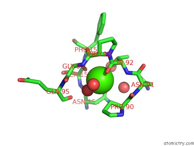
Mono view
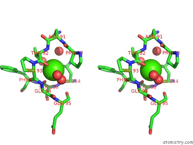
Stereo pair view
Mono view
Stereo pair view
A full contact list of Calcium with other atoms in the Ca binding
site number 1 of X-Ray Crystal Structure of Protein SPYM3_0169 From Streptococcus Pyogenes. Northeast Structural Genomics Consortium Target DR2. within 5.0Å range:
|
Calcium binding site 2 out of 2 in 1pg6
Go back to
Calcium binding site 2 out
of 2 in the X-Ray Crystal Structure of Protein SPYM3_0169 From Streptococcus Pyogenes. Northeast Structural Genomics Consortium Target DR2.
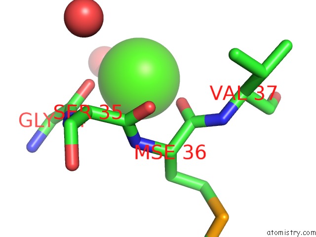
Mono view
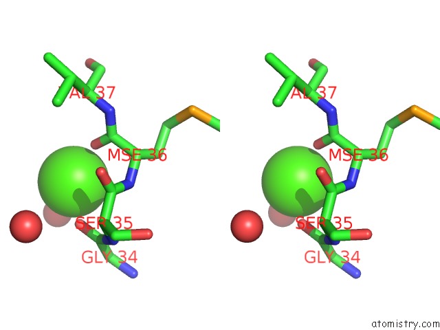
Stereo pair view
Mono view
Stereo pair view
A full contact list of Calcium with other atoms in the Ca binding
site number 2 of X-Ray Crystal Structure of Protein SPYM3_0169 From Streptococcus Pyogenes. Northeast Structural Genomics Consortium Target DR2. within 5.0Å range:
|
Reference:
A.Kuzin,
I.Lee,
W.Edstrom,
R.Xiao,
T.Acton,
B.Rost,
G.Montelione,
J.Hunt,
L.Tong.
X-Ray Structure of Hypothetical Protein SPYM3_0169 From Streptococcus Pyogenes To Be Published 2003.
Page generated: Thu Jul 11 13:45:04 2024
Last articles
Zn in 9J0NZn in 9J0O
Zn in 9J0P
Zn in 9FJX
Zn in 9EKB
Zn in 9C0F
Zn in 9CAH
Zn in 9CH0
Zn in 9CH3
Zn in 9CH1