Calcium »
PDB 1pig-1px2 »
1pjx »
Calcium in PDB 1pjx: 0.85 Angstrom Structure of Squid Ganglion Dfpase
Enzymatic activity of 0.85 Angstrom Structure of Squid Ganglion Dfpase
All present enzymatic activity of 0.85 Angstrom Structure of Squid Ganglion Dfpase:
3.1.8.2;
3.1.8.2;
Protein crystallography data
The structure of 0.85 Angstrom Structure of Squid Ganglion Dfpase, PDB code: 1pjx
was solved by
J.Koepke,
H.Rueterjans,
C.Luecke,
G.Fritzsch,
with X-Ray Crystallography technique. A brief refinement statistics is given in the table below:
| Resolution Low / High (Å) | 10.00 / 0.85 |
| Space group | P 21 21 21 |
| Cell size a, b, c (Å), α, β, γ (°) | 43.114, 81.849, 86.467, 90.00, 90.00, 90.00 |
| R / Rfree (%) | 12.1 / 12.8 |
Calcium Binding Sites:
The binding sites of Calcium atom in the 0.85 Angstrom Structure of Squid Ganglion Dfpase
(pdb code 1pjx). This binding sites where shown within
5.0 Angstroms radius around Calcium atom.
In total 2 binding sites of Calcium where determined in the 0.85 Angstrom Structure of Squid Ganglion Dfpase, PDB code: 1pjx:
Jump to Calcium binding site number: 1; 2;
In total 2 binding sites of Calcium where determined in the 0.85 Angstrom Structure of Squid Ganglion Dfpase, PDB code: 1pjx:
Jump to Calcium binding site number: 1; 2;
Calcium binding site 1 out of 2 in 1pjx
Go back to
Calcium binding site 1 out
of 2 in the 0.85 Angstrom Structure of Squid Ganglion Dfpase
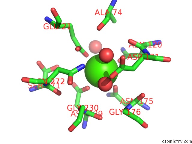
Mono view
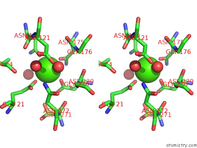
Stereo pair view
Mono view
Stereo pair view
A full contact list of Calcium with other atoms in the Ca binding
site number 1 of 0.85 Angstrom Structure of Squid Ganglion Dfpase within 5.0Å range:
|
Calcium binding site 2 out of 2 in 1pjx
Go back to
Calcium binding site 2 out
of 2 in the 0.85 Angstrom Structure of Squid Ganglion Dfpase
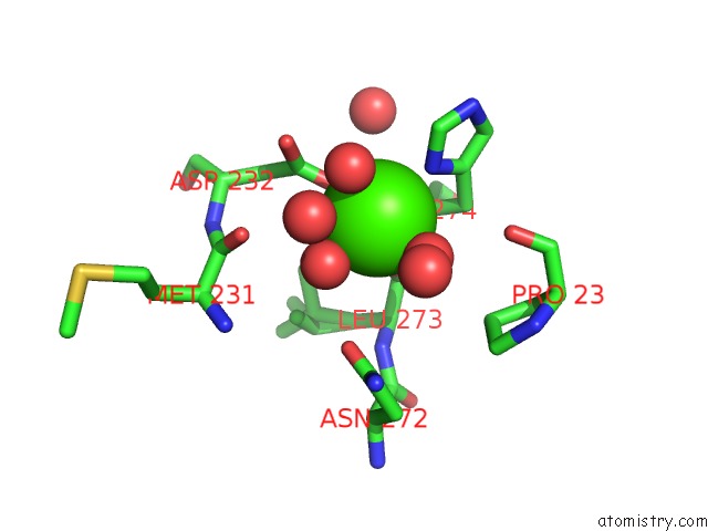
Mono view
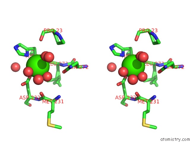
Stereo pair view
Mono view
Stereo pair view
A full contact list of Calcium with other atoms in the Ca binding
site number 2 of 0.85 Angstrom Structure of Squid Ganglion Dfpase within 5.0Å range:
|
Reference:
J.Koepke,
E.I.Scharff,
C.Lucke,
H.Ruterjans,
G.Fritzsch.
Statistical Analysis of Crystallographic Data Obtained From Squid Ganglion Dfpase at 0.85 A Resolution. Acta Crystallogr.,Sect.D V. 59 1744 2003.
ISSN: ISSN 0907-4449
PubMed: 14501113
DOI: 10.1107/S0907444903016135
Page generated: Thu Jul 11 13:48:10 2024
ISSN: ISSN 0907-4449
PubMed: 14501113
DOI: 10.1107/S0907444903016135
Last articles
Zn in 9J0NZn in 9J0O
Zn in 9J0P
Zn in 9FJX
Zn in 9EKB
Zn in 9C0F
Zn in 9CAH
Zn in 9CH0
Zn in 9CH3
Zn in 9CH1