Calcium »
PDB 1pig-1px2 »
1pmh »
Calcium in PDB 1pmh: Crystal Structure of Caldicellulosiruptor Saccharolyticus CBM27-1 in Complex with Mannohexaose
Enzymatic activity of Crystal Structure of Caldicellulosiruptor Saccharolyticus CBM27-1 in Complex with Mannohexaose
All present enzymatic activity of Crystal Structure of Caldicellulosiruptor Saccharolyticus CBM27-1 in Complex with Mannohexaose:
3.2.1.78;
3.2.1.78;
Protein crystallography data
The structure of Crystal Structure of Caldicellulosiruptor Saccharolyticus CBM27-1 in Complex with Mannohexaose, PDB code: 1pmh
was solved by
Y.Roske,
A.Sunna,
U.Heinemann,
with X-Ray Crystallography technique. A brief refinement statistics is given in the table below:
| Resolution Low / High (Å) | 23.00 / 1.06 |
| Space group | P 21 21 21 |
| Cell size a, b, c (Å), α, β, γ (°) | 38.281, 45.700, 110.113, 90.00, 90.00, 90.00 |
| R / Rfree (%) | 14.1 / 17.3 |
Calcium Binding Sites:
The binding sites of Calcium atom in the Crystal Structure of Caldicellulosiruptor Saccharolyticus CBM27-1 in Complex with Mannohexaose
(pdb code 1pmh). This binding sites where shown within
5.0 Angstroms radius around Calcium atom.
In total only one binding site of Calcium was determined in the Crystal Structure of Caldicellulosiruptor Saccharolyticus CBM27-1 in Complex with Mannohexaose, PDB code: 1pmh:
In total only one binding site of Calcium was determined in the Crystal Structure of Caldicellulosiruptor Saccharolyticus CBM27-1 in Complex with Mannohexaose, PDB code: 1pmh:
Calcium binding site 1 out of 1 in 1pmh
Go back to
Calcium binding site 1 out
of 1 in the Crystal Structure of Caldicellulosiruptor Saccharolyticus CBM27-1 in Complex with Mannohexaose
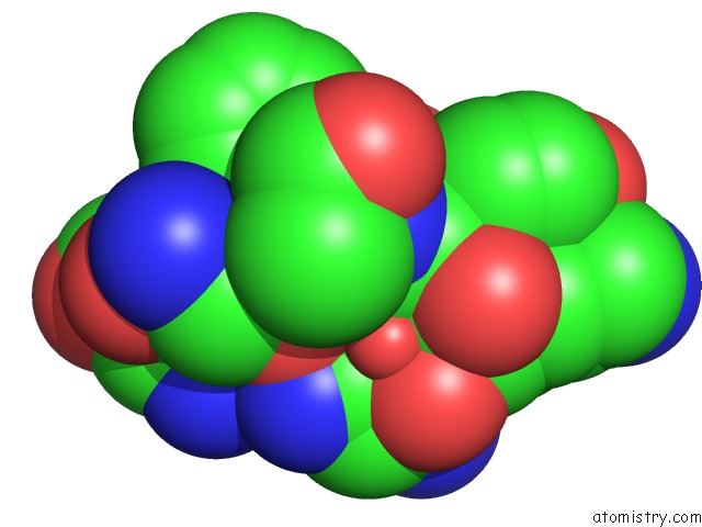
Mono view
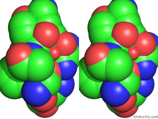
Stereo pair view
Mono view
Stereo pair view
A full contact list of Calcium with other atoms in the Ca binding
site number 1 of Crystal Structure of Caldicellulosiruptor Saccharolyticus CBM27-1 in Complex with Mannohexaose within 5.0Å range:
|
Reference:
Y.Roske,
A.Sunna,
W.Pfeil,
U.Heinemann.
High-Resolution Crystal Structures of Caldicellulosiruptor Strain RT8B.4 Carbohydrate-Binding Module CBM27-1 and Its Complex with Mannohexaose. J.Mol.Biol. V. 340 543 2004.
ISSN: ISSN 0022-2836
PubMed: 15210353
DOI: 10.1016/J.JMB.2004.04.072
Page generated: Thu Jul 11 13:48:44 2024
ISSN: ISSN 0022-2836
PubMed: 15210353
DOI: 10.1016/J.JMB.2004.04.072
Last articles
Zn in 9J0NZn in 9J0O
Zn in 9J0P
Zn in 9FJX
Zn in 9EKB
Zn in 9C0F
Zn in 9CAH
Zn in 9CH0
Zn in 9CH3
Zn in 9CH1