Calcium »
PDB 1pig-1px2 »
1pob »
Calcium in PDB 1pob: Crystal Structure of Cobra-Venom Phospholipase A2 in A Complex with A Transition-State Analogue
Enzymatic activity of Crystal Structure of Cobra-Venom Phospholipase A2 in A Complex with A Transition-State Analogue
All present enzymatic activity of Crystal Structure of Cobra-Venom Phospholipase A2 in A Complex with A Transition-State Analogue:
3.1.1.4;
3.1.1.4;
Protein crystallography data
The structure of Crystal Structure of Cobra-Venom Phospholipase A2 in A Complex with A Transition-State Analogue, PDB code: 1pob
was solved by
S.P.White,
D.L.Scott,
Z.Otwinowski,
P.B.Sigler,
with X-Ray Crystallography technique. A brief refinement statistics is given in the table below:
| Resolution Low / High (Å) | N/A / 2.00 |
| Space group | C 2 2 21 |
| Cell size a, b, c (Å), α, β, γ (°) | 34.600, 73.500, 181.600, 90.00, 90.00, 90.00 |
| R / Rfree (%) | n/a / n/a |
Calcium Binding Sites:
The binding sites of Calcium atom in the Crystal Structure of Cobra-Venom Phospholipase A2 in A Complex with A Transition-State Analogue
(pdb code 1pob). This binding sites where shown within
5.0 Angstroms radius around Calcium atom.
In total 4 binding sites of Calcium where determined in the Crystal Structure of Cobra-Venom Phospholipase A2 in A Complex with A Transition-State Analogue, PDB code: 1pob:
Jump to Calcium binding site number: 1; 2; 3; 4;
In total 4 binding sites of Calcium where determined in the Crystal Structure of Cobra-Venom Phospholipase A2 in A Complex with A Transition-State Analogue, PDB code: 1pob:
Jump to Calcium binding site number: 1; 2; 3; 4;
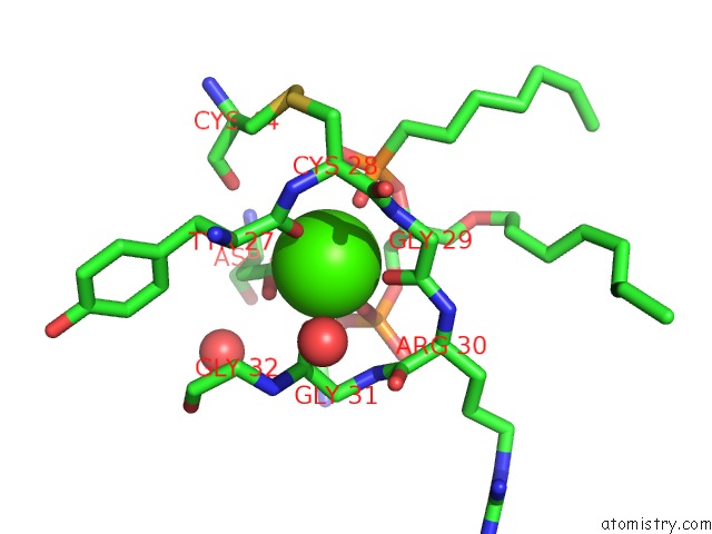
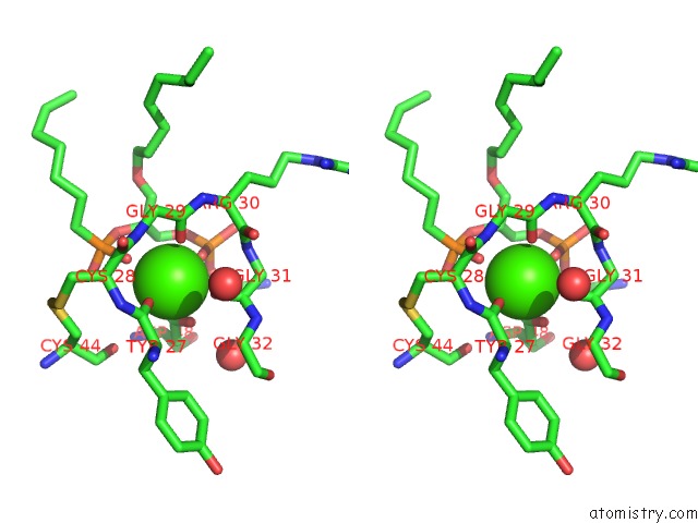
Calcium binding site 1 out of 4 in 1pob
Go back to
Calcium binding site 1 out
of 4 in the Crystal Structure of Cobra-Venom Phospholipase A2 in A Complex with A Transition-State Analogue
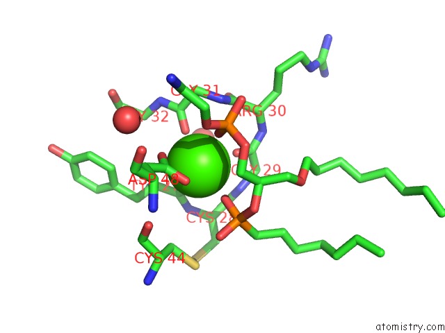
Mono view
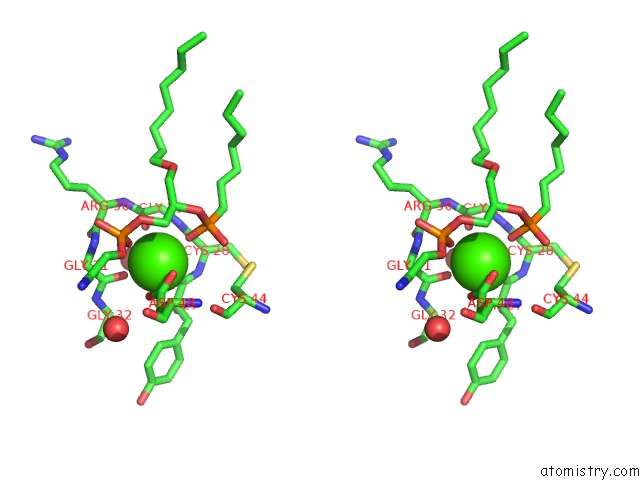
Stereo pair view
Mono view
Stereo pair view
A full contact list of Calcium with other atoms in the Ca binding
site number 1 of Crystal Structure of Cobra-Venom Phospholipase A2 in A Complex with A Transition-State Analogue within 5.0Å range:
|
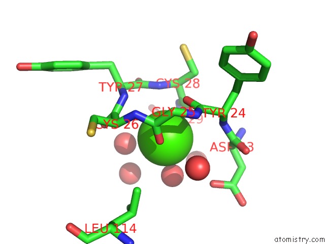
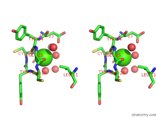
Calcium binding site 2 out of 4 in 1pob
Go back to
Calcium binding site 2 out
of 4 in the Crystal Structure of Cobra-Venom Phospholipase A2 in A Complex with A Transition-State Analogue
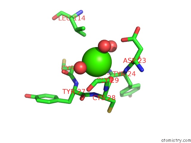
Mono view
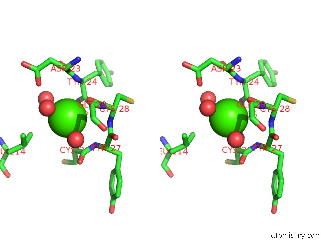
Stereo pair view
Mono view
Stereo pair view
A full contact list of Calcium with other atoms in the Ca binding
site number 2 of Crystal Structure of Cobra-Venom Phospholipase A2 in A Complex with A Transition-State Analogue within 5.0Å range:
|
Calcium binding site 3 out of 4 in 1pob
Go back to
Calcium binding site 3 out
of 4 in the Crystal Structure of Cobra-Venom Phospholipase A2 in A Complex with A Transition-State Analogue
Mono view
Stereo pair view
Mono view
Stereo pair view
A full contact list of Calcium with other atoms in the Ca binding
site number 3 of Crystal Structure of Cobra-Venom Phospholipase A2 in A Complex with A Transition-State Analogue within 5.0Å range:
|
Calcium binding site 4 out of 4 in 1pob
Go back to
Calcium binding site 4 out
of 4 in the Crystal Structure of Cobra-Venom Phospholipase A2 in A Complex with A Transition-State Analogue
Mono view
Stereo pair view
Mono view
Stereo pair view
A full contact list of Calcium with other atoms in the Ca binding
site number 4 of Crystal Structure of Cobra-Venom Phospholipase A2 in A Complex with A Transition-State Analogue within 5.0Å range:
|
Reference:
S.P.White,
D.L.Scott,
Z.Otwinowski,
M.H.Gelb,
P.B.Sigler.
Crystal Structure of Cobra-Venom Phospholipase A2 in A Complex with A Transition-State Analogue. Science V. 250 1560 1990.
ISSN: ISSN 0036-8075
PubMed: 2274787
Page generated: Thu Jul 11 13:49:58 2024
ISSN: ISSN 0036-8075
PubMed: 2274787
Last articles
Zn in 9J0NZn in 9J0O
Zn in 9J0P
Zn in 9FJX
Zn in 9EKB
Zn in 9C0F
Zn in 9CAH
Zn in 9CH0
Zn in 9CH3
Zn in 9CH1