Calcium »
PDB 1pig-1px2 »
1pvb »
Calcium in PDB 1pvb: X-Ray Structure of A New Crystal Form of Pike 4.10 Parvalbumin
Protein crystallography data
The structure of X-Ray Structure of A New Crystal Form of Pike 4.10 Parvalbumin, PDB code: 1pvb
was solved by
J.P.Declercq,
B.Tinant,
J.Parello,
with X-Ray Crystallography technique. A brief refinement statistics is given in the table below:
| Resolution Low / High (Å) | 8.00 / 1.75 |
| Space group | P 21 21 21 |
| Cell size a, b, c (Å), α, β, γ (°) | 51.840, 49.950, 34.960, 90.00, 90.00, 90.00 |
| R / Rfree (%) | 16.8 / n/a |
Calcium Binding Sites:
The binding sites of Calcium atom in the X-Ray Structure of A New Crystal Form of Pike 4.10 Parvalbumin
(pdb code 1pvb). This binding sites where shown within
5.0 Angstroms radius around Calcium atom.
In total 2 binding sites of Calcium where determined in the X-Ray Structure of A New Crystal Form of Pike 4.10 Parvalbumin, PDB code: 1pvb:
Jump to Calcium binding site number: 1; 2;
In total 2 binding sites of Calcium where determined in the X-Ray Structure of A New Crystal Form of Pike 4.10 Parvalbumin, PDB code: 1pvb:
Jump to Calcium binding site number: 1; 2;
Calcium binding site 1 out of 2 in 1pvb
Go back to
Calcium binding site 1 out
of 2 in the X-Ray Structure of A New Crystal Form of Pike 4.10 Parvalbumin
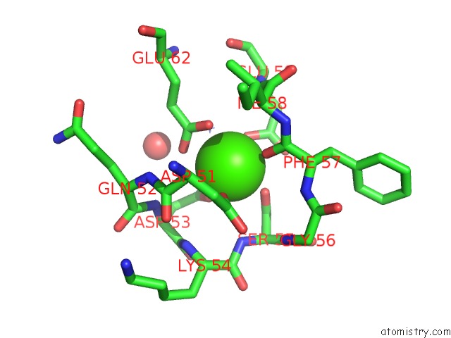
Mono view
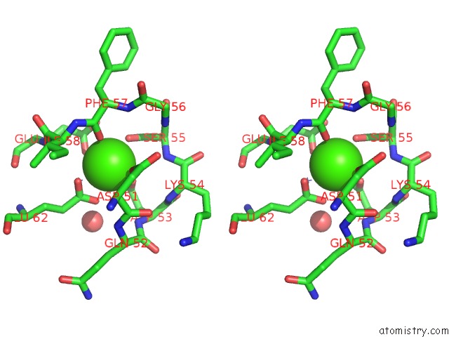
Stereo pair view
Mono view
Stereo pair view
A full contact list of Calcium with other atoms in the Ca binding
site number 1 of X-Ray Structure of A New Crystal Form of Pike 4.10 Parvalbumin within 5.0Å range:
|
Calcium binding site 2 out of 2 in 1pvb
Go back to
Calcium binding site 2 out
of 2 in the X-Ray Structure of A New Crystal Form of Pike 4.10 Parvalbumin
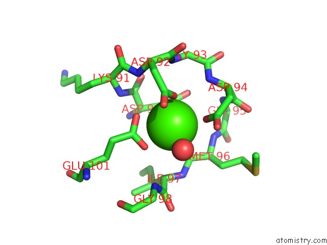
Mono view
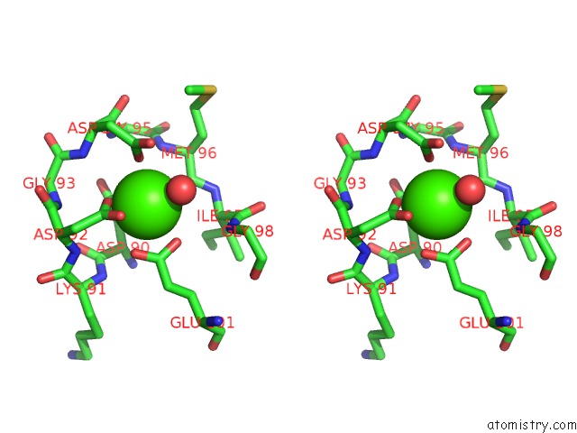
Stereo pair view
Mono view
Stereo pair view
A full contact list of Calcium with other atoms in the Ca binding
site number 2 of X-Ray Structure of A New Crystal Form of Pike 4.10 Parvalbumin within 5.0Å range:
|
Reference:
J.P.Declercq,
B.Tinant,
J.Parello.
X-Ray Structure of A New Crystal Form of Pike 4.10 Beta Parvalbumin. Acta Crystallogr.,Sect.D V. 52 165 1996.
ISSN: ISSN 0907-4449
PubMed: 15299738
DOI: 10.1107/S0907444995010006
Page generated: Thu Jul 11 13:53:16 2024
ISSN: ISSN 0907-4449
PubMed: 15299738
DOI: 10.1107/S0907444995010006
Last articles
Zn in 9J0NZn in 9J0O
Zn in 9J0P
Zn in 9FJX
Zn in 9EKB
Zn in 9C0F
Zn in 9CAH
Zn in 9CH0
Zn in 9CH3
Zn in 9CH1