Calcium »
PDB 1pig-1px2 »
1px2 »
Calcium in PDB 1px2: Crystal Structure of Rat Synapsin I C Domain Complexed to Ca.Atp (Form 1)
Protein crystallography data
The structure of Crystal Structure of Rat Synapsin I C Domain Complexed to Ca.Atp (Form 1), PDB code: 1px2
was solved by
C.A.Brautigam,
Y.Chelliah,
J.Deisenhofer,
with X-Ray Crystallography technique. A brief refinement statistics is given in the table below:
| Resolution Low / High (Å) | 20.00 / 2.23 |
| Space group | P 65 2 2 |
| Cell size a, b, c (Å), α, β, γ (°) | 96.000, 96.000, 305.800, 90.00, 90.00, 120.00 |
| R / Rfree (%) | 20.3 / 24 |
Calcium Binding Sites:
The binding sites of Calcium atom in the Crystal Structure of Rat Synapsin I C Domain Complexed to Ca.Atp (Form 1)
(pdb code 1px2). This binding sites where shown within
5.0 Angstroms radius around Calcium atom.
In total 2 binding sites of Calcium where determined in the Crystal Structure of Rat Synapsin I C Domain Complexed to Ca.Atp (Form 1), PDB code: 1px2:
Jump to Calcium binding site number: 1; 2;
In total 2 binding sites of Calcium where determined in the Crystal Structure of Rat Synapsin I C Domain Complexed to Ca.Atp (Form 1), PDB code: 1px2:
Jump to Calcium binding site number: 1; 2;
Calcium binding site 1 out of 2 in 1px2
Go back to
Calcium binding site 1 out
of 2 in the Crystal Structure of Rat Synapsin I C Domain Complexed to Ca.Atp (Form 1)
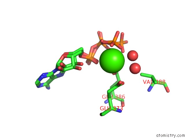
Mono view
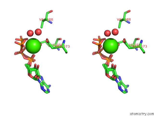
Stereo pair view
Mono view
Stereo pair view
A full contact list of Calcium with other atoms in the Ca binding
site number 1 of Crystal Structure of Rat Synapsin I C Domain Complexed to Ca.Atp (Form 1) within 5.0Å range:
|
Calcium binding site 2 out of 2 in 1px2
Go back to
Calcium binding site 2 out
of 2 in the Crystal Structure of Rat Synapsin I C Domain Complexed to Ca.Atp (Form 1)
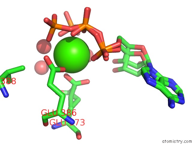
Mono view
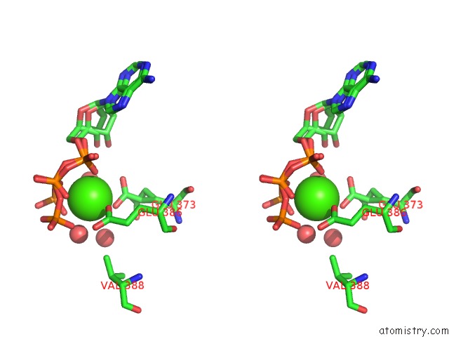
Stereo pair view
Mono view
Stereo pair view
A full contact list of Calcium with other atoms in the Ca binding
site number 2 of Crystal Structure of Rat Synapsin I C Domain Complexed to Ca.Atp (Form 1) within 5.0Å range:
|
Reference:
C.A.Brautigam,
Y.Chelliah,
J.Deisenhofer.
Tetramerization and Atp Binding By A Protein Comprising the A, B, and C Domains of Rat Synapsin I. J.Biol.Chem. V. 279 11948 2004.
ISSN: ISSN 0021-9258
PubMed: 14688264
DOI: 10.1074/JBC.M312015200
Page generated: Mon Jul 7 18:19:23 2025
ISSN: ISSN 0021-9258
PubMed: 14688264
DOI: 10.1074/JBC.M312015200
Last articles
Cl in 5L5SCl in 5L5T
Cl in 5L5R
Cl in 5L5Q
Cl in 5L5P
Cl in 5L5O
Cl in 5L5J
Cl in 5L5I
Cl in 5L5F
Cl in 5L5H