Calcium »
PDB 1px7-1qcp »
1qaf »
Calcium in PDB 1qaf: The Active Site Base Controls Cofactor Reactivity in Escherichia Coli Amine Oxidase : X-Ray Crystallographic Studies with Mutational Variants
Enzymatic activity of The Active Site Base Controls Cofactor Reactivity in Escherichia Coli Amine Oxidase : X-Ray Crystallographic Studies with Mutational Variants
All present enzymatic activity of The Active Site Base Controls Cofactor Reactivity in Escherichia Coli Amine Oxidase : X-Ray Crystallographic Studies with Mutational Variants:
1.4.3.4;
1.4.3.4;
Protein crystallography data
The structure of The Active Site Base Controls Cofactor Reactivity in Escherichia Coli Amine Oxidase : X-Ray Crystallographic Studies with Mutational Variants, PDB code: 1qaf
was solved by
J.M.Murray,
C.M.Wilmot,
C.G.Saysell,
J.Jaeger,
P.F.Knowles,
S.E.Phillips,
M.J.Mcpherson,
with X-Ray Crystallography technique. A brief refinement statistics is given in the table below:
| Resolution Low / High (Å) | 20.00 / 2.20 |
| Space group | P 21 21 21 |
| Cell size a, b, c (Å), α, β, γ (°) | 135.200, 167.000, 79.930, 90.00, 90.00, 90.00 |
| R / Rfree (%) | 17.6 / 24.4 |
Other elements in 1qaf:
The structure of The Active Site Base Controls Cofactor Reactivity in Escherichia Coli Amine Oxidase : X-Ray Crystallographic Studies with Mutational Variants also contains other interesting chemical elements:
| Copper | (Cu) | 2 atoms |
Calcium Binding Sites:
The binding sites of Calcium atom in the The Active Site Base Controls Cofactor Reactivity in Escherichia Coli Amine Oxidase : X-Ray Crystallographic Studies with Mutational Variants
(pdb code 1qaf). This binding sites where shown within
5.0 Angstroms radius around Calcium atom.
In total 4 binding sites of Calcium where determined in the The Active Site Base Controls Cofactor Reactivity in Escherichia Coli Amine Oxidase : X-Ray Crystallographic Studies with Mutational Variants, PDB code: 1qaf:
Jump to Calcium binding site number: 1; 2; 3; 4;
In total 4 binding sites of Calcium where determined in the The Active Site Base Controls Cofactor Reactivity in Escherichia Coli Amine Oxidase : X-Ray Crystallographic Studies with Mutational Variants, PDB code: 1qaf:
Jump to Calcium binding site number: 1; 2; 3; 4;
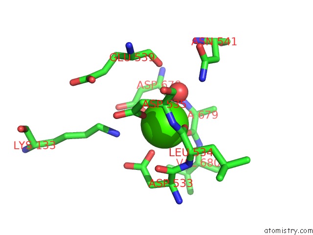
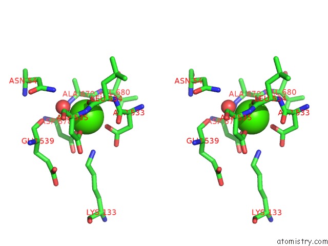
Calcium binding site 1 out of 4 in 1qaf
Go back to
Calcium binding site 1 out
of 4 in the The Active Site Base Controls Cofactor Reactivity in Escherichia Coli Amine Oxidase : X-Ray Crystallographic Studies with Mutational Variants
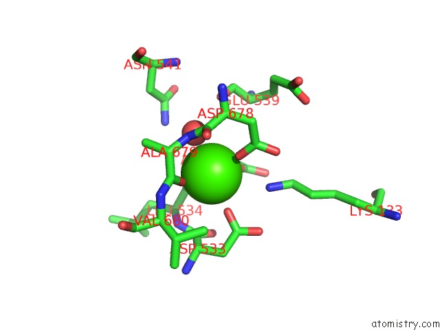
Mono view
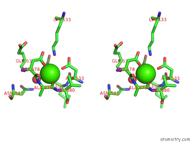
Stereo pair view
Mono view
Stereo pair view
A full contact list of Calcium with other atoms in the Ca binding
site number 1 of The Active Site Base Controls Cofactor Reactivity in Escherichia Coli Amine Oxidase : X-Ray Crystallographic Studies with Mutational Variants within 5.0Å range:
|
Calcium binding site 2 out of 4 in 1qaf
Go back to
Calcium binding site 2 out
of 4 in the The Active Site Base Controls Cofactor Reactivity in Escherichia Coli Amine Oxidase : X-Ray Crystallographic Studies with Mutational Variants
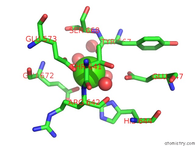
Mono view
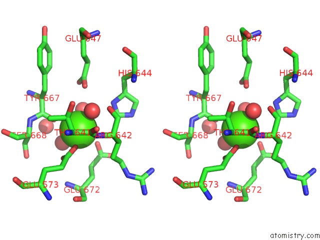
Stereo pair view
Mono view
Stereo pair view
A full contact list of Calcium with other atoms in the Ca binding
site number 2 of The Active Site Base Controls Cofactor Reactivity in Escherichia Coli Amine Oxidase : X-Ray Crystallographic Studies with Mutational Variants within 5.0Å range:
|
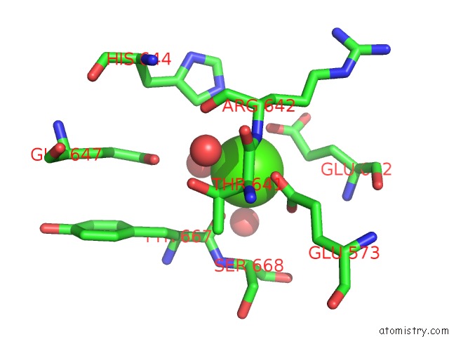
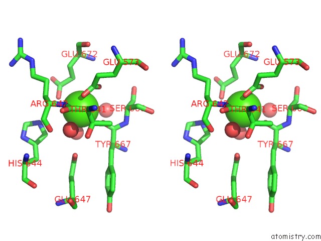
Calcium binding site 3 out of 4 in 1qaf
Go back to
Calcium binding site 3 out
of 4 in the The Active Site Base Controls Cofactor Reactivity in Escherichia Coli Amine Oxidase : X-Ray Crystallographic Studies with Mutational Variants
Mono view
Stereo pair view
Mono view
Stereo pair view
A full contact list of Calcium with other atoms in the Ca binding
site number 3 of The Active Site Base Controls Cofactor Reactivity in Escherichia Coli Amine Oxidase : X-Ray Crystallographic Studies with Mutational Variants within 5.0Å range:
|
Calcium binding site 4 out of 4 in 1qaf
Go back to
Calcium binding site 4 out
of 4 in the The Active Site Base Controls Cofactor Reactivity in Escherichia Coli Amine Oxidase : X-Ray Crystallographic Studies with Mutational Variants
Mono view
Stereo pair view
Mono view
Stereo pair view
A full contact list of Calcium with other atoms in the Ca binding
site number 4 of The Active Site Base Controls Cofactor Reactivity in Escherichia Coli Amine Oxidase : X-Ray Crystallographic Studies with Mutational Variants within 5.0Å range:
|
Reference:
J.M.Murray,
C.G.Saysell,
C.M.Wilmot,
W.S.Tambyrajah,
J.Jaeger,
P.F.Knowles,
S.E.Phillips,
M.J.Mcpherson.
The Active Site Base Controls Cofactor Reactivity in Escherichia Coli Amine Oxidase: X-Ray Crystallographic Studies with Mutational Variants. Biochemistry V. 38 8217 1999.
ISSN: ISSN 0006-2960
PubMed: 10387067
DOI: 10.1021/BI9900469
Page generated: Thu Jul 11 14:17:17 2024
ISSN: ISSN 0006-2960
PubMed: 10387067
DOI: 10.1021/BI9900469
Last articles
Zn in 9MJ5Zn in 9HNW
Zn in 9G0L
Zn in 9FNE
Zn in 9DZN
Zn in 9E0I
Zn in 9D32
Zn in 9DAK
Zn in 8ZXC
Zn in 8ZUF