Calcium »
PDB 1px7-1qcp »
1qco »
Calcium in PDB 1qco: Crystal Structure of Fumarylacetoacetate Hydrolase Complexed with Fumarate and Acetoacetate
Enzymatic activity of Crystal Structure of Fumarylacetoacetate Hydrolase Complexed with Fumarate and Acetoacetate
All present enzymatic activity of Crystal Structure of Fumarylacetoacetate Hydrolase Complexed with Fumarate and Acetoacetate:
3.7.1.2;
3.7.1.2;
Protein crystallography data
The structure of Crystal Structure of Fumarylacetoacetate Hydrolase Complexed with Fumarate and Acetoacetate, PDB code: 1qco
was solved by
D.E.Timm,
H.A.Mueller,
P.Bhanumoorthy,
J.M.Harp,
G.J.Bunick,
with X-Ray Crystallography technique. A brief refinement statistics is given in the table below:
| Resolution Low / High (Å) | 27.90 / 1.90 |
| Space group | P 1 21 1 |
| Cell size a, b, c (Å), α, β, γ (°) | 64.640, 110.940, 67.810, 90.00, 102.52, 90.00 |
| R / Rfree (%) | 15.9 / 19.2 |
Other elements in 1qco:
The structure of Crystal Structure of Fumarylacetoacetate Hydrolase Complexed with Fumarate and Acetoacetate also contains other interesting chemical elements:
| Nickel | (Ni) | 5 atoms |
Calcium Binding Sites:
The binding sites of Calcium atom in the Crystal Structure of Fumarylacetoacetate Hydrolase Complexed with Fumarate and Acetoacetate
(pdb code 1qco). This binding sites where shown within
5.0 Angstroms radius around Calcium atom.
In total 2 binding sites of Calcium where determined in the Crystal Structure of Fumarylacetoacetate Hydrolase Complexed with Fumarate and Acetoacetate, PDB code: 1qco:
Jump to Calcium binding site number: 1; 2;
In total 2 binding sites of Calcium where determined in the Crystal Structure of Fumarylacetoacetate Hydrolase Complexed with Fumarate and Acetoacetate, PDB code: 1qco:
Jump to Calcium binding site number: 1; 2;
Calcium binding site 1 out of 2 in 1qco
Go back to
Calcium binding site 1 out
of 2 in the Crystal Structure of Fumarylacetoacetate Hydrolase Complexed with Fumarate and Acetoacetate
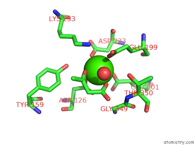
Mono view
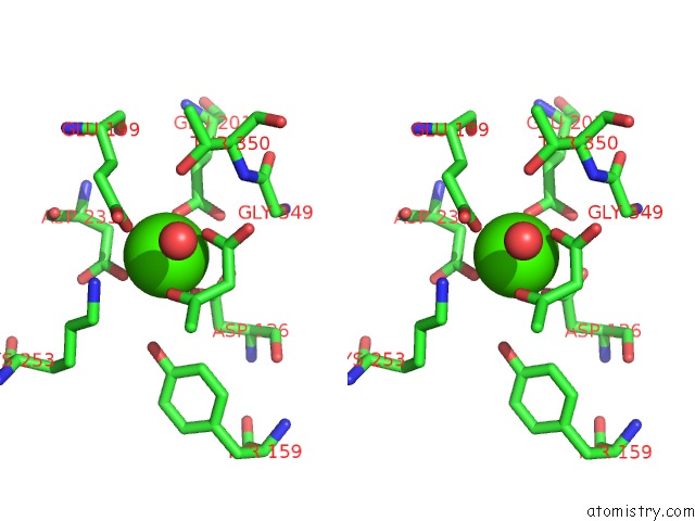
Stereo pair view
Mono view
Stereo pair view
A full contact list of Calcium with other atoms in the Ca binding
site number 1 of Crystal Structure of Fumarylacetoacetate Hydrolase Complexed with Fumarate and Acetoacetate within 5.0Å range:
|
Calcium binding site 2 out of 2 in 1qco
Go back to
Calcium binding site 2 out
of 2 in the Crystal Structure of Fumarylacetoacetate Hydrolase Complexed with Fumarate and Acetoacetate
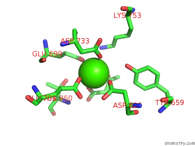
Mono view
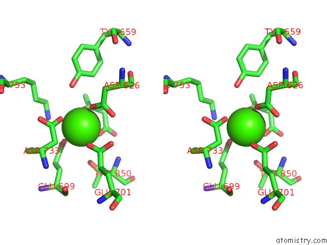
Stereo pair view
Mono view
Stereo pair view
A full contact list of Calcium with other atoms in the Ca binding
site number 2 of Crystal Structure of Fumarylacetoacetate Hydrolase Complexed with Fumarate and Acetoacetate within 5.0Å range:
|
Reference:
D.E.Timm,
H.A.Mueller,
P.Bhanumoorthy,
J.M.Harp,
G.J.Bunick.
Crystal Structure and Mechanism of A Carbon-Carbon Bond Hydrolase. Structure Fold.Des. V. 7 1023 1999.
ISSN: ISSN 0969-2126
PubMed: 10508789
DOI: 10.1016/S0969-2126(99)80170-1
Page generated: Mon Jul 7 23:10:44 2025
ISSN: ISSN 0969-2126
PubMed: 10508789
DOI: 10.1016/S0969-2126(99)80170-1
Last articles
F in 7NTHF in 7NTI
F in 7NPC
F in 7NRG
F in 7NR5
F in 7NQS
F in 7NOS
F in 7NP5
F in 7NDV
F in 7NP6