Calcium »
PDB 1qd6-1qlk »
1qe5 »
Calcium in PDB 1qe5: Purine Nucleoside Phosphorylase From Cellulomonas Sp. in Complex with Phosphate
Enzymatic activity of Purine Nucleoside Phosphorylase From Cellulomonas Sp. in Complex with Phosphate
All present enzymatic activity of Purine Nucleoside Phosphorylase From Cellulomonas Sp. in Complex with Phosphate:
2.4.2.1;
2.4.2.1;
Protein crystallography data
The structure of Purine Nucleoside Phosphorylase From Cellulomonas Sp. in Complex with Phosphate, PDB code: 1qe5
was solved by
J.Tebbe,
A.Bzowska,
B.Wielgus-Kutrowska,
W.Schroeder,
Z.Kazimierczuk,
D.Shugar,
W.Saenger,
G.Koellner,
with X-Ray Crystallography technique. A brief refinement statistics is given in the table below:
| Resolution Low / High (Å) | 28.00 / 2.20 |
| Space group | P 21 21 21 |
| Cell size a, b, c (Å), α, β, γ (°) | 64.120, 108.900, 119.300, 90.00, 90.00, 90.00 |
| R / Rfree (%) | 20 / 26 |
Calcium Binding Sites:
The binding sites of Calcium atom in the Purine Nucleoside Phosphorylase From Cellulomonas Sp. in Complex with Phosphate
(pdb code 1qe5). This binding sites where shown within
5.0 Angstroms radius around Calcium atom.
In total only one binding site of Calcium was determined in the Purine Nucleoside Phosphorylase From Cellulomonas Sp. in Complex with Phosphate, PDB code: 1qe5:
In total only one binding site of Calcium was determined in the Purine Nucleoside Phosphorylase From Cellulomonas Sp. in Complex with Phosphate, PDB code: 1qe5:
Calcium binding site 1 out of 1 in 1qe5
Go back to
Calcium binding site 1 out
of 1 in the Purine Nucleoside Phosphorylase From Cellulomonas Sp. in Complex with Phosphate
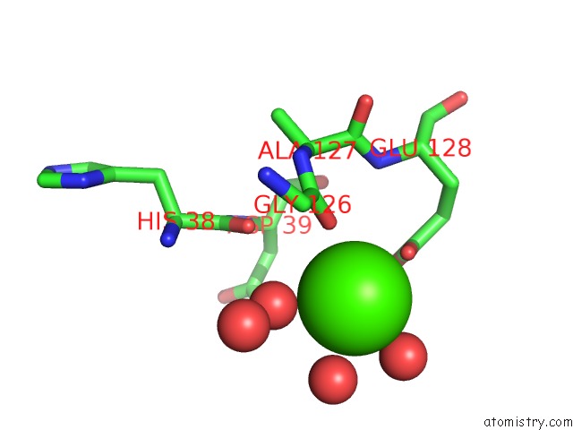
Mono view
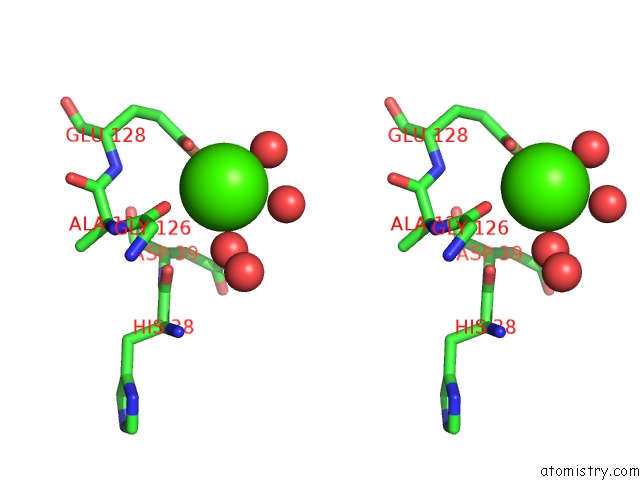
Stereo pair view
Mono view
Stereo pair view
A full contact list of Calcium with other atoms in the Ca binding
site number 1 of Purine Nucleoside Phosphorylase From Cellulomonas Sp. in Complex with Phosphate within 5.0Å range:
|
Reference:
J.Tebbe,
A.Bzowska,
B.Wielgus-Kutrowska,
W.Schroder,
Z.Kazimierczuk,
D.Shugar,
W.Saenger,
G.Koellner.
Crystal Structure of the Purine Nucleoside Phosphorylase (Pnp) From Cellulomonas Sp. and Its Implication For the Mechanism of Trimeric Pnps. J.Mol.Biol. V. 294 1239 1999.
ISSN: ISSN 0022-2836
PubMed: 10600382
DOI: 10.1006/JMBI.1999.3327
Page generated: Thu Jul 11 21:42:24 2024
ISSN: ISSN 0022-2836
PubMed: 10600382
DOI: 10.1006/JMBI.1999.3327
Last articles
Zn in 9J0NZn in 9J0O
Zn in 9J0P
Zn in 9FJX
Zn in 9EKB
Zn in 9C0F
Zn in 9CAH
Zn in 9CH0
Zn in 9CH3
Zn in 9CH1