Calcium »
PDB 1qd6-1qlk »
1qf3 »
Calcium in PDB 1qf3: Peanut Lectin Complexed with Methyl-Beta-Galactose
Protein crystallography data
The structure of Peanut Lectin Complexed with Methyl-Beta-Galactose, PDB code: 1qf3
was solved by
R.Ravishankar,
K.Suguna,
A.Surolia,
M.Vijayan,
with X-Ray Crystallography technique. A brief refinement statistics is given in the table below:
| Resolution Low / High (Å) | 10.00 / 2.80 |
| Space group | P 21 21 2 |
| Cell size a, b, c (Å), α, β, γ (°) | 128.953, 126.683, 77.284, 90.00, 90.00, 90.00 |
| R / Rfree (%) | 19.5 / 24.7 |
Other elements in 1qf3:
The structure of Peanut Lectin Complexed with Methyl-Beta-Galactose also contains other interesting chemical elements:
| Manganese | (Mn) | 4 atoms |
Calcium Binding Sites:
The binding sites of Calcium atom in the Peanut Lectin Complexed with Methyl-Beta-Galactose
(pdb code 1qf3). This binding sites where shown within
5.0 Angstroms radius around Calcium atom.
In total 4 binding sites of Calcium where determined in the Peanut Lectin Complexed with Methyl-Beta-Galactose, PDB code: 1qf3:
Jump to Calcium binding site number: 1; 2; 3; 4;
In total 4 binding sites of Calcium where determined in the Peanut Lectin Complexed with Methyl-Beta-Galactose, PDB code: 1qf3:
Jump to Calcium binding site number: 1; 2; 3; 4;
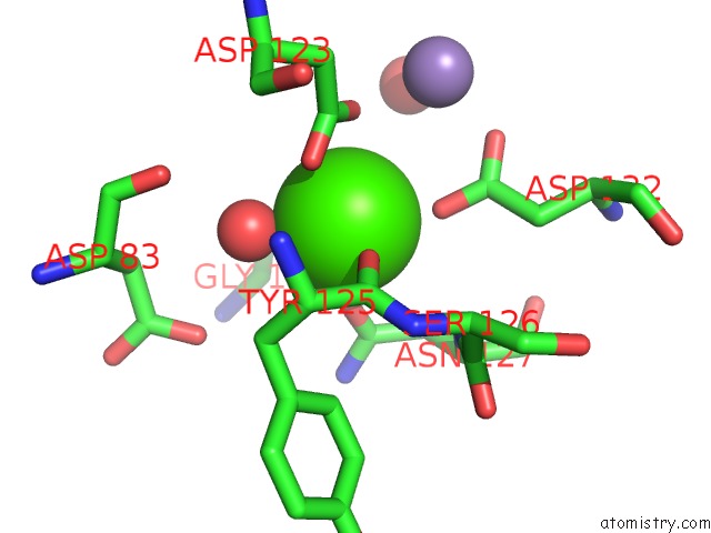
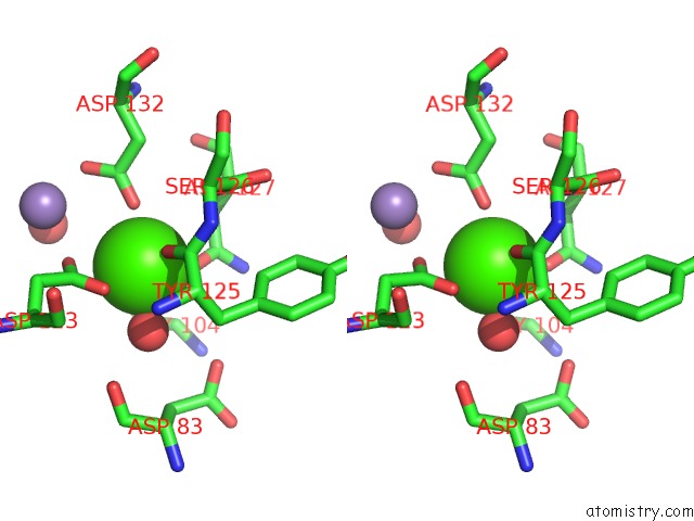
Calcium binding site 1 out of 4 in 1qf3
Go back to
Calcium binding site 1 out
of 4 in the Peanut Lectin Complexed with Methyl-Beta-Galactose
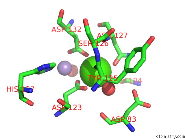
Mono view
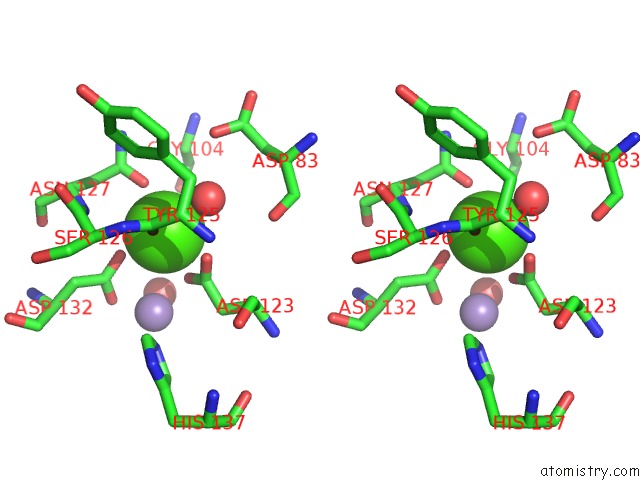
Stereo pair view
Mono view
Stereo pair view
A full contact list of Calcium with other atoms in the Ca binding
site number 1 of Peanut Lectin Complexed with Methyl-Beta-Galactose within 5.0Å range:
|
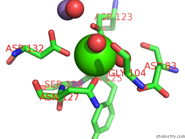
Calcium binding site 2 out of 4 in 1qf3
Go back to
Calcium binding site 2 out
of 4 in the Peanut Lectin Complexed with Methyl-Beta-Galactose
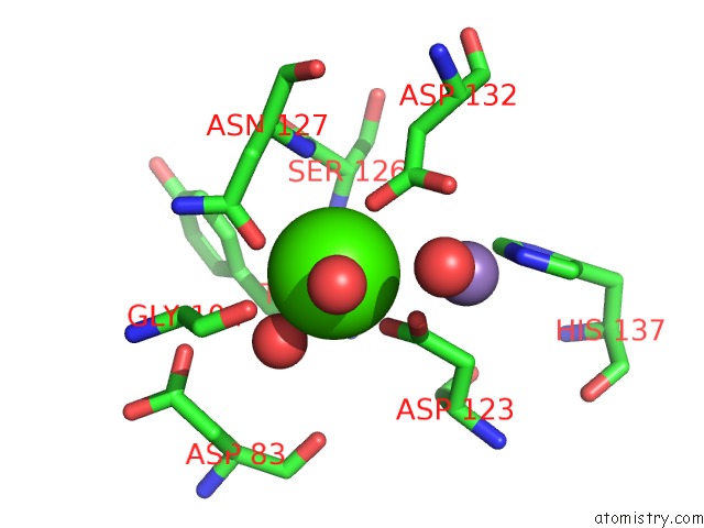
Mono view
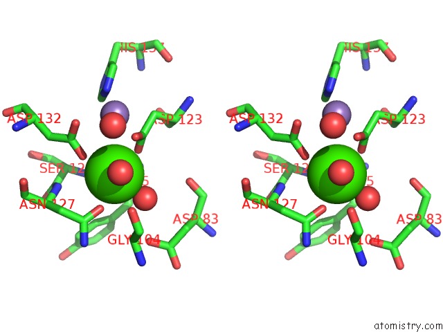
Stereo pair view
Mono view
Stereo pair view
A full contact list of Calcium with other atoms in the Ca binding
site number 2 of Peanut Lectin Complexed with Methyl-Beta-Galactose within 5.0Å range:
|
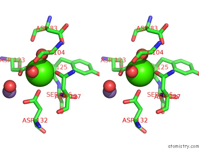
Calcium binding site 3 out of 4 in 1qf3
Go back to
Calcium binding site 3 out
of 4 in the Peanut Lectin Complexed with Methyl-Beta-Galactose
Mono view
Stereo pair view
Mono view
Stereo pair view
A full contact list of Calcium with other atoms in the Ca binding
site number 3 of Peanut Lectin Complexed with Methyl-Beta-Galactose within 5.0Å range:
|
Calcium binding site 4 out of 4 in 1qf3
Go back to
Calcium binding site 4 out
of 4 in the Peanut Lectin Complexed with Methyl-Beta-Galactose
Mono view
Stereo pair view
Mono view
Stereo pair view
A full contact list of Calcium with other atoms in the Ca binding
site number 4 of Peanut Lectin Complexed with Methyl-Beta-Galactose within 5.0Å range:
|
Reference:
R.Ravishankar,
K.Suguna,
A.Surolia,
M.Vijayan.
Structures of the Complexes of Peanut Lectin with Methyl-Beta-Galactose and N-Acetyllactosamine and A Comparative Study of Carbohydrate Binding in Gal/Galnac-Specific Legume Lectins. Acta Crystallogr.,Sect.D V. 55 1375 1999.
ISSN: ISSN 0907-4449
PubMed: 10417405
DOI: 10.1107/S0907444999006587
Page generated: Tue Jul 8 01:16:40 2025
ISSN: ISSN 0907-4449
PubMed: 10417405
DOI: 10.1107/S0907444999006587
Last articles
Cl in 5L0TCl in 5L0S
Cl in 5L0R
Cl in 5L0E
Cl in 5L0L
Cl in 5L0B
Cl in 5KXN
Cl in 5KZ6
Cl in 5L01
Cl in 5KWE