Calcium »
PDB 1qls-1ra1 »
1qz5 »
Calcium in PDB 1qz5: Structure of Rabbit Actin in Complex with Kabiramide C
Protein crystallography data
The structure of Structure of Rabbit Actin in Complex with Kabiramide C, PDB code: 1qz5
was solved by
V.A.Klenchin,
J.S.Allingham,
R.King,
J.Tanaka,
G.Marriott,
I.Rayment,
with X-Ray Crystallography technique. A brief refinement statistics is given in the table below:
| Resolution Low / High (Å) | 30.00 / 1.45 |
| Space group | P 21 21 21 |
| Cell size a, b, c (Å), α, β, γ (°) | 69.919, 70.617, 75.130, 90.00, 90.00, 90.00 |
| R / Rfree (%) | 16.9 / 18.7 |
Calcium Binding Sites:
The binding sites of Calcium atom in the Structure of Rabbit Actin in Complex with Kabiramide C
(pdb code 1qz5). This binding sites where shown within
5.0 Angstroms radius around Calcium atom.
In total only one binding site of Calcium was determined in the Structure of Rabbit Actin in Complex with Kabiramide C, PDB code: 1qz5:
In total only one binding site of Calcium was determined in the Structure of Rabbit Actin in Complex with Kabiramide C, PDB code: 1qz5:
Calcium binding site 1 out of 1 in 1qz5
Go back to
Calcium binding site 1 out
of 1 in the Structure of Rabbit Actin in Complex with Kabiramide C
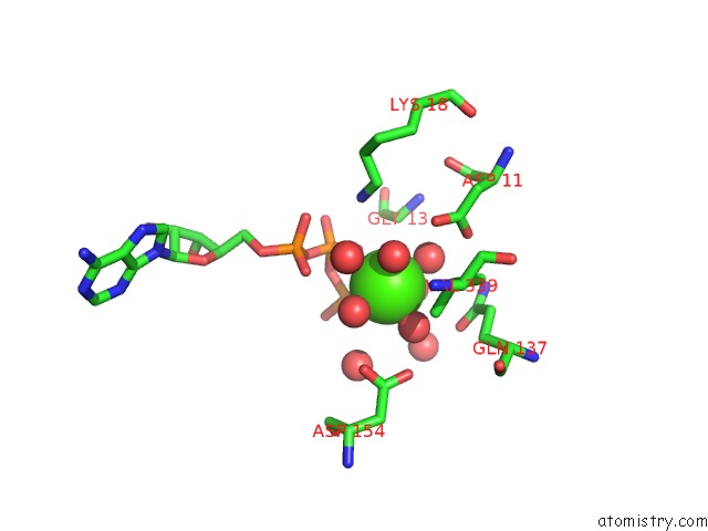
Mono view
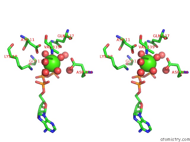
Stereo pair view
Mono view
Stereo pair view
A full contact list of Calcium with other atoms in the Ca binding
site number 1 of Structure of Rabbit Actin in Complex with Kabiramide C within 5.0Å range:
|
Reference:
V.A.Klenchin,
J.S.Allingham,
R.King,
J.Tanaka,
G.Marriott,
I.Rayment.
Trisoxazole Macrolide Toxins Mimic the Binding of Actin-Capping Proteins to Actin Nat.Struct.Biol. V. 10 1058 2003.
ISSN: ISSN 1072-8368
PubMed: 14578936
DOI: 10.1038/NSB1006
Page generated: Thu Jul 11 22:00:36 2024
ISSN: ISSN 1072-8368
PubMed: 14578936
DOI: 10.1038/NSB1006
Last articles
Zn in 9J0NZn in 9J0O
Zn in 9J0P
Zn in 9FJX
Zn in 9EKB
Zn in 9C0F
Zn in 9CAH
Zn in 9CH0
Zn in 9CH3
Zn in 9CH1