Calcium in PDB 1r0r: 1.1 Angstrom Resolution Structure of the Complex Between the Protein Inhibitor, OMTKY3, and the Serine Protease, Subtilisin Carlsberg
Enzymatic activity of 1.1 Angstrom Resolution Structure of the Complex Between the Protein Inhibitor, OMTKY3, and the Serine Protease, Subtilisin Carlsberg
All present enzymatic activity of 1.1 Angstrom Resolution Structure of the Complex Between the Protein Inhibitor, OMTKY3, and the Serine Protease, Subtilisin Carlsberg:
3.4.21.62;
Protein crystallography data
The structure of 1.1 Angstrom Resolution Structure of the Complex Between the Protein Inhibitor, OMTKY3, and the Serine Protease, Subtilisin Carlsberg, PDB code: 1r0r
was solved by
J.R.Horn,
S.Ramaswamy,
K.P.Murphy,
with X-Ray Crystallography technique. A brief refinement statistics is given in the table below:
Resolution Low / High (Å)
|
15.25 /
1.10
|
Space group
|
C 2 2 21
|
Cell size a, b, c (Å), α, β, γ (°)
|
62.329,
70.810,
127.476,
90.00,
90.00,
90.00
|
R / Rfree (%)
|
15.8 /
18.4
|
Calcium Binding Sites:
The binding sites of Calcium atom in the 1.1 Angstrom Resolution Structure of the Complex Between the Protein Inhibitor, OMTKY3, and the Serine Protease, Subtilisin Carlsberg
(pdb code 1r0r). This binding sites where shown within
5.0 Angstroms radius around Calcium atom.
In total 6 binding sites of Calcium where determined in the
1.1 Angstrom Resolution Structure of the Complex Between the Protein Inhibitor, OMTKY3, and the Serine Protease, Subtilisin Carlsberg, PDB code: 1r0r:
Jump to Calcium binding site number:
1;
2;
3;
4;
5;
6;
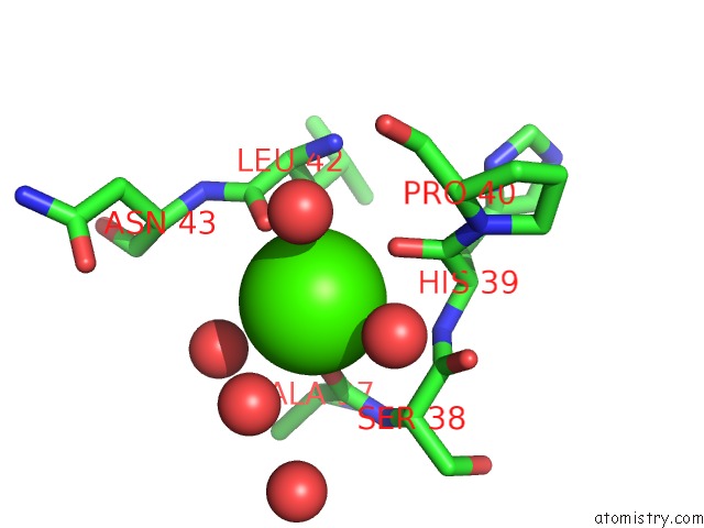
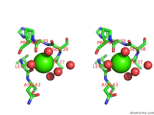
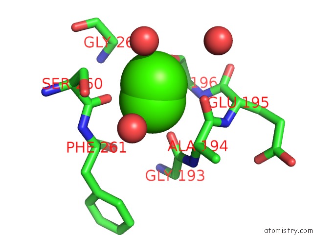
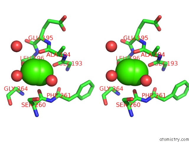
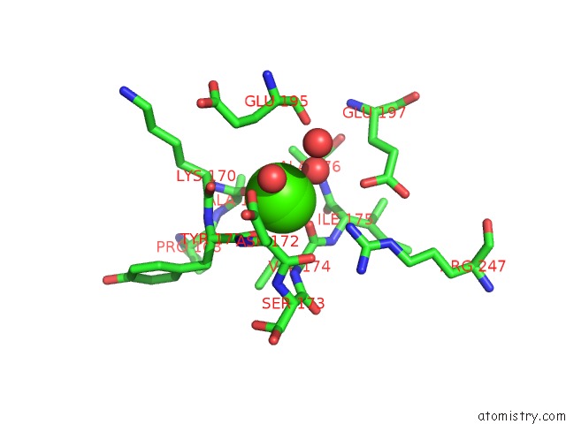
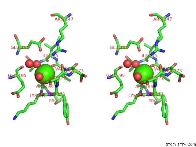
Calcium binding site 1 out
of 6 in 1r0r
Go back to
Calcium Binding Sites List in 1r0r
Calcium binding site 1 out
of 6 in the 1.1 Angstrom Resolution Structure of the Complex Between the Protein Inhibitor, OMTKY3, and the Serine Protease, Subtilisin Carlsberg
 Mono view
 Stereo pair view
|
A full contact list of Calcium with other atoms in the Ca binding
site number 1 of 1.1 Angstrom Resolution Structure of the Complex Between the Protein Inhibitor, OMTKY3, and the Serine Protease, Subtilisin Carlsberg within 5.0Å range:
probe
|
atom
|
residue
|
distance (Å)
|
B
|
Occ
|
E:Ca301
b:22.7
occ:1.00
|
O
|
E:HIS39
|
2.6
|
11.6
|
1.0
|
O
|
E:LEU42
|
2.7
|
12.7
|
1.0
|
O
|
E:ALA37
|
2.8
|
11.8
|
1.0
|
O
|
E:HOH4096
|
2.8
|
33.8
|
1.0
|
O
|
E:HOH4097
|
2.9
|
29.8
|
1.0
|
O
|
E:HOH4126
|
2.9
|
27.2
|
1.0
|
O
|
E:HOH4009
|
3.4
|
20.3
|
1.0
|
C
|
E:HIS39
|
3.7
|
11.9
|
1.0
|
C
|
E:LEU42
|
3.8
|
13.2
|
1.0
|
C
|
E:SER38
|
3.9
|
12.8
|
1.0
|
C
|
E:ALA37
|
4.0
|
11.9
|
1.0
|
O
|
E:SER38
|
4.0
|
12.8
|
1.0
|
N
|
E:HIS39
|
4.1
|
12.2
|
1.0
|
CA
|
E:SER38
|
4.1
|
12.7
|
1.0
|
CA
|
E:PRO40
|
4.5
|
12.9
|
1.0
|
CA
|
E:HIS39
|
4.5
|
12.4
|
1.0
|
N
|
E:PRO40
|
4.5
|
12.6
|
1.0
|
N
|
E:LEU42
|
4.5
|
12.8
|
1.0
|
N
|
E:SER38
|
4.5
|
12.8
|
1.0
|
C
|
E:PRO40
|
4.6
|
12.6
|
1.0
|
CA
|
E:LEU42
|
4.7
|
12.6
|
1.0
|
O
|
E:PRO40
|
4.7
|
13.2
|
1.0
|
N
|
E:ASN43
|
4.8
|
13.3
|
1.0
|
CA
|
E:ASN43
|
4.8
|
13.5
|
1.0
|
O
|
E:HOH4066
|
4.8
|
23.8
|
1.0
|
|
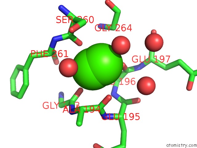
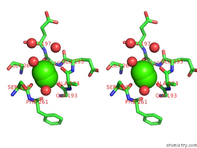
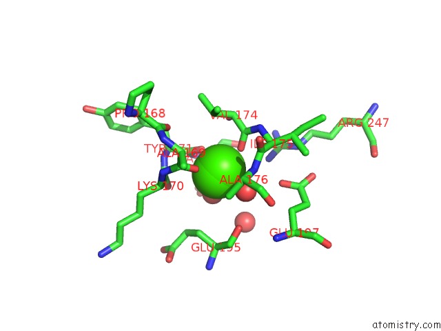
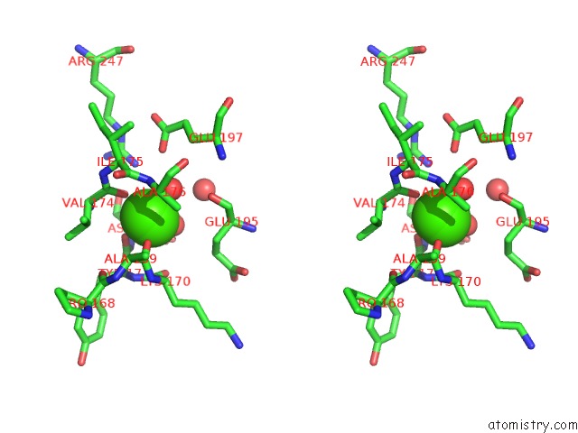
Calcium binding site 2 out
of 6 in 1r0r
Go back to
Calcium Binding Sites List in 1r0r
Calcium binding site 2 out
of 6 in the 1.1 Angstrom Resolution Structure of the Complex Between the Protein Inhibitor, OMTKY3, and the Serine Protease, Subtilisin Carlsberg
 Mono view
 Stereo pair view
|
A full contact list of Calcium with other atoms in the Ca binding
site number 2 of 1.1 Angstrom Resolution Structure of the Complex Between the Protein Inhibitor, OMTKY3, and the Serine Protease, Subtilisin Carlsberg within 5.0Å range:
probe
|
atom
|
residue
|
distance (Å)
|
B
|
Occ
|
E:Ca302
b:19.1
occ:1.00
|
O
|
E:LEU75
|
2.3
|
11.6
|
1.0
|
O
|
E:THR79
|
2.3
|
11.6
|
1.0
|
O
|
E:VAL81
|
2.3
|
12.8
|
1.0
|
OD1
|
E:ASN77
|
2.3
|
11.2
|
1.0
|
OE1
|
E:GLN2
|
2.4
|
12.6
|
1.0
|
OD1
|
E:ASP41
|
2.4
|
12.3
|
1.0
|
OD2
|
E:ASP41
|
2.5
|
11.8
|
1.0
|
CG
|
E:ASP41
|
2.8
|
12.4
|
1.0
|
CG
|
E:ASN77
|
3.4
|
12.6
|
1.0
|
CD
|
E:GLN2
|
3.4
|
12.9
|
1.0
|
C
|
E:LEU75
|
3.5
|
12.1
|
1.0
|
C
|
E:VAL81
|
3.5
|
13.3
|
1.0
|
C
|
E:THR79
|
3.5
|
11.9
|
1.0
|
N
|
E:VAL81
|
3.7
|
12.9
|
1.0
|
ND2
|
E:ASN77
|
3.9
|
14.2
|
1.0
|
CG
|
E:GLN2
|
3.9
|
14.0
|
1.0
|
N
|
E:ASN77
|
4.0
|
13.2
|
1.0
|
C
|
E:GLY80
|
4.1
|
13.5
|
1.0
|
CA
|
E:VAL81
|
4.2
|
13.4
|
1.0
|
N
|
E:LEU75
|
4.3
|
12.8
|
1.0
|
CA
|
E:LEU75
|
4.3
|
13.0
|
1.0
|
CB
|
E:ASP41
|
4.3
|
12.5
|
1.0
|
CA
|
E:GLY80
|
4.4
|
12.5
|
1.0
|
N
|
E:THR79
|
4.4
|
12.8
|
1.0
|
N
|
E:GLY80
|
4.4
|
12.8
|
1.0
|
N
|
E:ASP76
|
4.4
|
12.9
|
1.0
|
CA
|
E:THR79
|
4.5
|
12.6
|
1.0
|
N
|
E:LEU82
|
4.5
|
14.0
|
1.0
|
CG2
|
E:THR79
|
4.5
|
13.4
|
1.0
|
CB
|
E:ASN77
|
4.5
|
12.7
|
1.0
|
CB
|
E:GLN2
|
4.6
|
14.2
|
1.0
|
CA
|
E:ASP76
|
4.6
|
13.6
|
1.0
|
NE2
|
E:GLN2
|
4.6
|
14.1
|
1.0
|
CB
|
E:LEU75
|
4.6
|
13.1
|
1.0
|
CA
|
E:ASN77
|
4.6
|
13.3
|
1.0
|
C
|
E:ASN77
|
4.7
|
13.6
|
1.0
|
C
|
E:ASP76
|
4.7
|
13.6
|
1.0
|
O
|
E:ASN77
|
4.7
|
13.6
|
1.0
|
C
|
E:ALA74
|
4.8
|
12.3
|
1.0
|
CA
|
E:LEU82
|
4.8
|
13.8
|
1.0
|
O
|
E:GLY80
|
4.8
|
13.8
|
1.0
|
|
Calcium binding site 3 out
of 6 in 1r0r
Go back to
Calcium Binding Sites List in 1r0r
Calcium binding site 3 out
of 6 in the 1.1 Angstrom Resolution Structure of the Complex Between the Protein Inhibitor, OMTKY3, and the Serine Protease, Subtilisin Carlsberg
 Mono view
 Stereo pair view
|
A full contact list of Calcium with other atoms in the Ca binding
site number 3 of 1.1 Angstrom Resolution Structure of the Complex Between the Protein Inhibitor, OMTKY3, and the Serine Protease, Subtilisin Carlsberg within 5.0Å range:
probe
|
atom
|
residue
|
distance (Å)
|
B
|
Occ
|
E:Ca303
b:27.1
occ:0.50
|
CA
|
E:CA303
|
0.0
|
27.1
|
0.5
|
CA
|
E:CA303
|
0.9
|
27.0
|
0.5
|
O
|
E:GLY193
|
2.6
|
15.1
|
1.0
|
O
|
E:SER260
|
2.7
|
15.9
|
1.0
|
O
|
E:HOH4174
|
2.8
|
39.4
|
1.0
|
O
|
E:ALA194
|
2.9
|
15.8
|
1.0
|
O
|
E:LEU196
|
2.9
|
14.2
|
1.0
|
C
|
E:ALA194
|
3.3
|
14.7
|
1.0
|
O
|
E:HOH4286
|
3.4
|
43.0
|
1.0
|
C
|
E:SER260
|
3.4
|
17.0
|
1.0
|
CA
|
E:ALA194
|
3.5
|
15.6
|
1.0
|
C
|
E:GLY193
|
3.6
|
14.8
|
1.0
|
N
|
E:LEU196
|
3.7
|
14.1
|
1.0
|
C
|
E:LEU196
|
3.8
|
13.7
|
1.0
|
CB
|
E:LEU196
|
3.9
|
13.7
|
1.0
|
CA
|
E:LEU196
|
4.0
|
13.7
|
1.0
|
N
|
E:ALA194
|
4.0
|
15.6
|
1.0
|
CA
|
E:PHE261
|
4.1
|
16.0
|
1.0
|
N
|
E:PHE261
|
4.1
|
16.3
|
1.0
|
CB
|
E:SER260
|
4.2
|
18.5
|
1.0
|
N
|
E:GLU195
|
4.2
|
15.1
|
1.0
|
OG
|
E:SER260
|
4.3
|
20.4
|
1.0
|
CA
|
E:GLY264
|
4.3
|
14.5
|
1.0
|
CA
|
E:SER260
|
4.4
|
17.9
|
1.0
|
C
|
E:GLU195
|
4.4
|
14.3
|
1.0
|
N
|
E:GLY264
|
4.6
|
14.9
|
1.0
|
C
|
E:PHE261
|
4.6
|
15.7
|
1.0
|
O
|
E:PHE261
|
4.7
|
14.8
|
1.0
|
O
|
E:HOH4434
|
4.7
|
40.3
|
1.0
|
CA
|
E:GLU195
|
4.8
|
15.0
|
1.0
|
CG
|
E:LEU196
|
4.9
|
13.5
|
1.0
|
CA
|
E:GLY193
|
4.9
|
14.6
|
1.0
|
CB
|
E:ALA194
|
4.9
|
16.0
|
1.0
|
|
Calcium binding site 4 out
of 6 in 1r0r
Go back to
Calcium Binding Sites List in 1r0r
Calcium binding site 4 out
of 6 in the 1.1 Angstrom Resolution Structure of the Complex Between the Protein Inhibitor, OMTKY3, and the Serine Protease, Subtilisin Carlsberg
 Mono view
 Stereo pair view
|
A full contact list of Calcium with other atoms in the Ca binding
site number 4 of 1.1 Angstrom Resolution Structure of the Complex Between the Protein Inhibitor, OMTKY3, and the Serine Protease, Subtilisin Carlsberg within 5.0Å range:
probe
|
atom
|
residue
|
distance (Å)
|
B
|
Occ
|
E:Ca303
b:27.0
occ:0.50
|
CA
|
E:CA303
|
0.0
|
27.0
|
0.5
|
CA
|
E:CA303
|
0.9
|
27.1
|
0.5
|
O
|
E:LEU196
|
2.5
|
14.2
|
1.0
|
O
|
E:HOH4286
|
2.6
|
43.0
|
1.0
|
O
|
E:ALA194
|
2.9
|
15.8
|
1.0
|
O
|
E:SER260
|
2.9
|
15.9
|
1.0
|
O
|
E:HOH4174
|
3.2
|
39.4
|
1.0
|
O
|
E:GLY193
|
3.3
|
15.1
|
1.0
|
C
|
E:LEU196
|
3.5
|
13.7
|
1.0
|
C
|
E:ALA194
|
3.6
|
14.7
|
1.0
|
C
|
E:SER260
|
3.7
|
17.0
|
1.0
|
N
|
E:LEU196
|
3.8
|
14.1
|
1.0
|
CA
|
E:GLY264
|
3.9
|
14.5
|
1.0
|
CB
|
E:SER260
|
3.9
|
18.5
|
1.0
|
CA
|
E:LEU196
|
4.0
|
13.7
|
1.0
|
CA
|
E:ALA194
|
4.0
|
15.6
|
1.0
|
CB
|
E:LEU196
|
4.1
|
13.7
|
1.0
|
O
|
E:HOH4434
|
4.1
|
40.3
|
1.0
|
OG
|
E:SER260
|
4.2
|
20.4
|
1.0
|
CA
|
E:SER260
|
4.3
|
17.9
|
1.0
|
C
|
E:GLY193
|
4.4
|
14.8
|
1.0
|
C
|
E:GLU195
|
4.4
|
14.3
|
1.0
|
N
|
E:GLY264
|
4.5
|
14.9
|
1.0
|
N
|
E:GLU195
|
4.5
|
15.1
|
1.0
|
N
|
E:PHE261
|
4.5
|
16.3
|
1.0
|
N
|
E:GLU197
|
4.7
|
13.8
|
1.0
|
O
|
E:HOH4232
|
4.7
|
34.9
|
1.0
|
N
|
E:ALA194
|
4.7
|
15.6
|
1.0
|
CA
|
E:PHE261
|
4.7
|
16.0
|
1.0
|
CA
|
E:GLU195
|
4.9
|
15.0
|
1.0
|
C
|
E:GLY264
|
4.9
|
15.0
|
1.0
|
CG
|
E:LEU196
|
5.0
|
13.5
|
1.0
|
|
Calcium binding site 5 out
of 6 in 1r0r
Go back to
Calcium Binding Sites List in 1r0r
Calcium binding site 5 out
of 6 in the 1.1 Angstrom Resolution Structure of the Complex Between the Protein Inhibitor, OMTKY3, and the Serine Protease, Subtilisin Carlsberg
 Mono view
 Stereo pair view
|
A full contact list of Calcium with other atoms in the Ca binding
site number 5 of 1.1 Angstrom Resolution Structure of the Complex Between the Protein Inhibitor, OMTKY3, and the Serine Protease, Subtilisin Carlsberg within 5.0Å range:
probe
|
atom
|
residue
|
distance (Å)
|
B
|
Occ
|
E:Ca305
b:43.0
occ:0.50
|
CA
|
E:CA305
|
0.0
|
43.0
|
0.5
|
CA
|
E:CA305
|
0.3
|
22.8
|
0.5
|
O
|
E:VAL174
|
2.1
|
12.3
|
1.0
|
O
|
E:HOH4021
|
2.4
|
21.3
|
1.0
|
O
|
E:ALA169
|
2.4
|
13.3
|
1.0
|
O
|
E:TYR171
|
2.4
|
13.0
|
1.0
|
O
|
E:HOH4084
|
2.4
|
30.1
|
1.0
|
C
|
E:VAL174
|
3.2
|
12.8
|
1.0
|
C
|
E:TYR171
|
3.4
|
13.3
|
1.0
|
C
|
E:ALA169
|
3.6
|
14.2
|
1.0
|
C
|
E:LYS170
|
3.9
|
13.8
|
1.0
|
CA
|
E:VAL174
|
3.9
|
13.3
|
1.0
|
N
|
E:ALA176
|
3.9
|
13.1
|
1.0
|
N
|
E:VAL174
|
4.0
|
13.6
|
1.0
|
CB
|
E:VAL174
|
4.0
|
13.1
|
1.0
|
N
|
E:TYR171
|
4.1
|
13.9
|
1.0
|
O
|
E:LYS170
|
4.1
|
15.2
|
1.0
|
CA
|
E:ASP172
|
4.2
|
14.6
|
1.0
|
NH2
|
E:ARG247
|
4.2
|
17.9
|
1.0
|
N
|
E:ILE175
|
4.2
|
13.5
|
1.0
|
N
|
E:ASP172
|
4.2
|
14.2
|
1.0
|
CB
|
E:ALA176
|
4.3
|
13.3
|
1.0
|
CA
|
E:LYS170
|
4.3
|
14.6
|
1.0
|
CA
|
E:TYR171
|
4.4
|
13.4
|
1.0
|
N
|
E:LYS170
|
4.4
|
14.3
|
1.0
|
C
|
E:ASP172
|
4.4
|
13.3
|
1.0
|
O
|
E:GLU195
|
4.4
|
14.6
|
1.0
|
CA
|
E:ILE175
|
4.4
|
13.9
|
1.0
|
OE2
|
E:GLU197
|
4.4
|
16.1
|
1.0
|
C
|
E:ILE175
|
4.5
|
13.2
|
1.0
|
O
|
E:ASP172
|
4.5
|
13.4
|
1.0
|
CA
|
E:ALA169
|
4.6
|
13.8
|
1.0
|
CB
|
E:GLU195
|
4.6
|
15.3
|
1.0
|
CA
|
E:ALA176
|
4.7
|
13.1
|
1.0
|
CG1
|
E:VAL174
|
4.7
|
14.3
|
1.0
|
O
|
E:HOH4254
|
4.8
|
49.2
|
1.0
|
O
|
E:PRO168
|
5.0
|
13.2
|
1.0
|
N
|
E:SER173
|
5.0
|
13.5
|
1.0
|
|
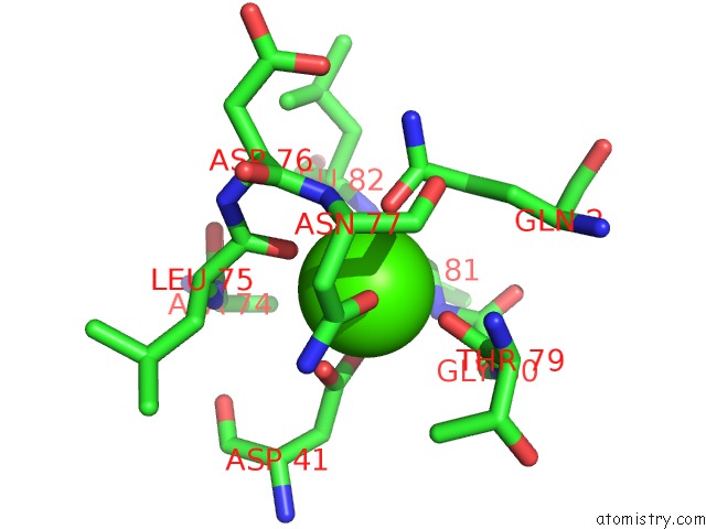
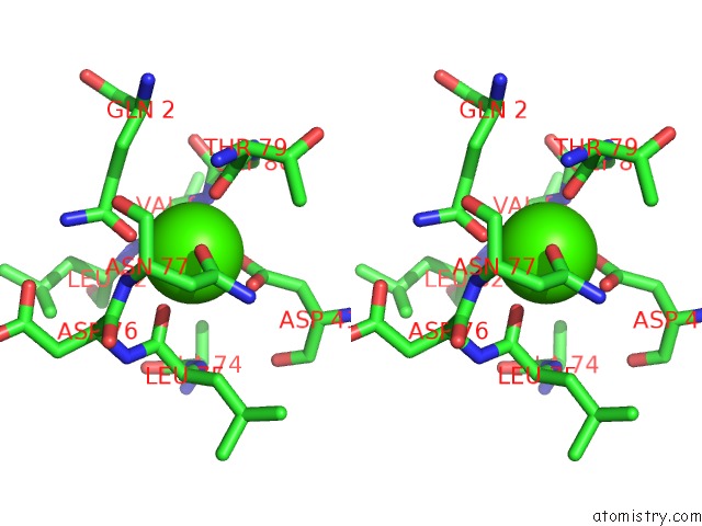
Calcium binding site 6 out
of 6 in 1r0r
Go back to
Calcium Binding Sites List in 1r0r
Calcium binding site 6 out
of 6 in the 1.1 Angstrom Resolution Structure of the Complex Between the Protein Inhibitor, OMTKY3, and the Serine Protease, Subtilisin Carlsberg
 Mono view
 Stereo pair view
|
A full contact list of Calcium with other atoms in the Ca binding
site number 6 of 1.1 Angstrom Resolution Structure of the Complex Between the Protein Inhibitor, OMTKY3, and the Serine Protease, Subtilisin Carlsberg within 5.0Å range:
probe
|
atom
|
residue
|
distance (Å)
|
B
|
Occ
|
E:Ca305
b:22.8
occ:0.50
|
CA
|
E:CA305
|
0.0
|
22.8
|
0.5
|
CA
|
E:CA305
|
0.3
|
43.0
|
0.5
|
O
|
E:HOH4084
|
2.2
|
30.1
|
1.0
|
O
|
E:ALA169
|
2.4
|
13.3
|
1.0
|
O
|
E:VAL174
|
2.4
|
12.3
|
1.0
|
O
|
E:HOH4021
|
2.4
|
21.3
|
1.0
|
O
|
E:TYR171
|
2.4
|
13.0
|
1.0
|
C
|
E:TYR171
|
3.4
|
13.3
|
1.0
|
C
|
E:VAL174
|
3.5
|
12.8
|
1.0
|
C
|
E:ALA169
|
3.5
|
14.2
|
1.0
|
C
|
E:LYS170
|
3.7
|
13.8
|
1.0
|
O
|
E:LYS170
|
3.9
|
15.2
|
1.0
|
N
|
E:TYR171
|
3.9
|
13.9
|
1.0
|
CA
|
E:LYS170
|
4.1
|
14.6
|
1.0
|
CA
|
E:ASP172
|
4.1
|
14.6
|
1.0
|
N
|
E:ASP172
|
4.1
|
14.2
|
1.0
|
N
|
E:ALA176
|
4.1
|
13.1
|
1.0
|
N
|
E:VAL174
|
4.2
|
13.6
|
1.0
|
CA
|
E:VAL174
|
4.2
|
13.3
|
1.0
|
CB
|
E:VAL174
|
4.3
|
13.1
|
1.0
|
N
|
E:LYS170
|
4.3
|
14.3
|
1.0
|
CA
|
E:TYR171
|
4.3
|
13.4
|
1.0
|
CB
|
E:ALA176
|
4.3
|
13.3
|
1.0
|
O
|
E:GLU195
|
4.3
|
14.6
|
1.0
|
CB
|
E:GLU195
|
4.4
|
15.3
|
1.0
|
NH2
|
E:ARG247
|
4.4
|
17.9
|
1.0
|
C
|
E:ASP172
|
4.5
|
13.3
|
1.0
|
N
|
E:ILE175
|
4.5
|
13.5
|
1.0
|
OE2
|
E:GLU197
|
4.6
|
16.1
|
1.0
|
CA
|
E:ALA169
|
4.6
|
13.8
|
1.0
|
O
|
E:ASP172
|
4.6
|
13.4
|
1.0
|
O
|
E:HOH4254
|
4.7
|
49.2
|
1.0
|
C
|
E:ILE175
|
4.7
|
13.2
|
1.0
|
CA
|
E:ILE175
|
4.7
|
13.9
|
1.0
|
CA
|
E:ALA176
|
4.8
|
13.1
|
1.0
|
CG
|
E:GLU195
|
4.8
|
16.6
|
1.0
|
OD2
|
E:ASP172
|
4.9
|
22.7
|
1.0
|
CG1
|
E:VAL174
|
5.0
|
14.3
|
1.0
|
O
|
E:PRO168
|
5.0
|
13.2
|
1.0
|
|
Reference:
J.R.Horn,
S.Ramaswamy,
K.P.Murphy.
Structure and Energetics of Protein-Protein Interactions: the Role of Conformational Heterogeneity in OMTKY3 Binding to Serine Proteases J.Mol.Biol. V. 331 497 2003.
ISSN: ISSN 0022-2836
PubMed: 12888355
DOI: 10.1016/S0022-2836(03)00783-6
Page generated: Thu Jul 11 22:01:31 2024
|