Calcium »
PDB 1rc8-1rpk »
1rf1 »
Calcium in PDB 1rf1: Crystal Structure of Fragment D of GAMMAE132A Fibrinogen with the Peptide Ligand Gly-His-Arg-Pro-Amide
Protein crystallography data
The structure of Crystal Structure of Fragment D of GAMMAE132A Fibrinogen with the Peptide Ligand Gly-His-Arg-Pro-Amide, PDB code: 1rf1
was solved by
M.S.Kostelansky,
O.V.Gorkun,
S.T.Lord,
with X-Ray Crystallography technique. A brief refinement statistics is given in the table below:
| Resolution Low / High (Å) | 17.97 / 2.53 |
| Space group | P 21 21 21 |
| Cell size a, b, c (Å), α, β, γ (°) | 89.714, 94.683, 228.198, 90.00, 90.00, 90.00 |
| R / Rfree (%) | 23.4 / 28.1 |
Calcium Binding Sites:
The binding sites of Calcium atom in the Crystal Structure of Fragment D of GAMMAE132A Fibrinogen with the Peptide Ligand Gly-His-Arg-Pro-Amide
(pdb code 1rf1). This binding sites where shown within
5.0 Angstroms radius around Calcium atom.
In total 4 binding sites of Calcium where determined in the Crystal Structure of Fragment D of GAMMAE132A Fibrinogen with the Peptide Ligand Gly-His-Arg-Pro-Amide, PDB code: 1rf1:
Jump to Calcium binding site number: 1; 2; 3; 4;
In total 4 binding sites of Calcium where determined in the Crystal Structure of Fragment D of GAMMAE132A Fibrinogen with the Peptide Ligand Gly-His-Arg-Pro-Amide, PDB code: 1rf1:
Jump to Calcium binding site number: 1; 2; 3; 4;
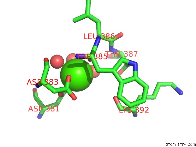
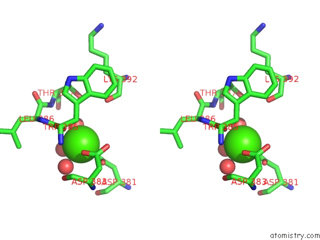
Calcium binding site 1 out of 4 in 1rf1
Go back to
Calcium binding site 1 out
of 4 in the Crystal Structure of Fragment D of GAMMAE132A Fibrinogen with the Peptide Ligand Gly-His-Arg-Pro-Amide
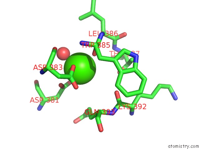
Mono view
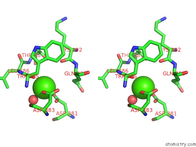
Stereo pair view
Mono view
Stereo pair view
A full contact list of Calcium with other atoms in the Ca binding
site number 1 of Crystal Structure of Fragment D of GAMMAE132A Fibrinogen with the Peptide Ligand Gly-His-Arg-Pro-Amide within 5.0Å range:
|
Calcium binding site 2 out of 4 in 1rf1
Go back to
Calcium binding site 2 out
of 4 in the Crystal Structure of Fragment D of GAMMAE132A Fibrinogen with the Peptide Ligand Gly-His-Arg-Pro-Amide
Mono view
Stereo pair view
Mono view
Stereo pair view
A full contact list of Calcium with other atoms in the Ca binding
site number 2 of Crystal Structure of Fragment D of GAMMAE132A Fibrinogen with the Peptide Ligand Gly-His-Arg-Pro-Amide within 5.0Å range:
|
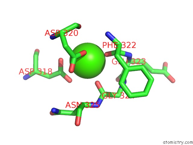
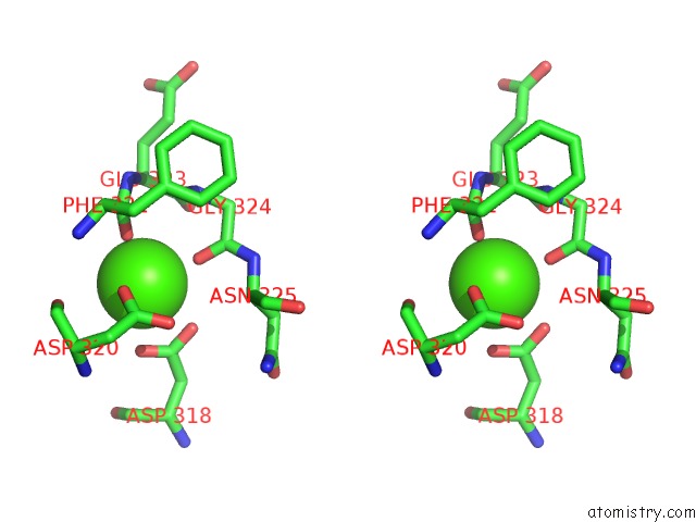
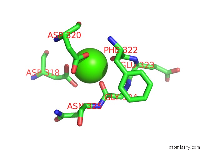
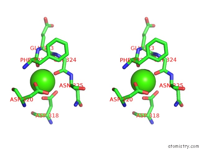
Calcium binding site 3 out of 4 in 1rf1
Go back to
Calcium binding site 3 out
of 4 in the Crystal Structure of Fragment D of GAMMAE132A Fibrinogen with the Peptide Ligand Gly-His-Arg-Pro-Amide
Mono view
Stereo pair view
Mono view
Stereo pair view
A full contact list of Calcium with other atoms in the Ca binding
site number 3 of Crystal Structure of Fragment D of GAMMAE132A Fibrinogen with the Peptide Ligand Gly-His-Arg-Pro-Amide within 5.0Å range:
|
Calcium binding site 4 out of 4 in 1rf1
Go back to
Calcium binding site 4 out
of 4 in the Crystal Structure of Fragment D of GAMMAE132A Fibrinogen with the Peptide Ligand Gly-His-Arg-Pro-Amide
Mono view
Stereo pair view
Mono view
Stereo pair view
A full contact list of Calcium with other atoms in the Ca binding
site number 4 of Crystal Structure of Fragment D of GAMMAE132A Fibrinogen with the Peptide Ligand Gly-His-Arg-Pro-Amide within 5.0Å range:
|
Reference:
M.S.Kostelansky,
K.C.Lounes,
L.F.Ping,
S.K.Dickerson,
O.V.Gorkun,
S.T.Lord.
Calcium-Binding Site BETA2, Adjacent to the "B" Polymerization Site, Modulates Lateral Aggregation of Protofibrils During Fibrin Polymerization. Biochemistry V. 43 2475 2004.
ISSN: ISSN 0006-2960
PubMed: 14992585
DOI: 10.1021/BI0359978
Page generated: Thu Jul 11 22:10:00 2024
ISSN: ISSN 0006-2960
PubMed: 14992585
DOI: 10.1021/BI0359978
Last articles
Zn in 9J0NZn in 9J0O
Zn in 9J0P
Zn in 9FJX
Zn in 9EKB
Zn in 9C0F
Zn in 9CAH
Zn in 9CH0
Zn in 9CH3
Zn in 9CH1