Calcium »
PDB 1rc8-1rpk »
1rir »
Calcium in PDB 1rir: Crystal Structure of Meso-Tetrasulphonatophenylporphyrin in Complex with Peanut Lectin.
Protein crystallography data
The structure of Crystal Structure of Meso-Tetrasulphonatophenylporphyrin in Complex with Peanut Lectin., PDB code: 1rir
was solved by
M.Goel,
K.J.Kaur,
B.G.Maiya,
M.J.Swamy,
D.M.Salunke,
with X-Ray Crystallography technique. A brief refinement statistics is given in the table below:
| Resolution Low / High (Å) | 100.00 / 2.90 |
| Space group | P 32 |
| Cell size a, b, c (Å), α, β, γ (°) | 94.800, 94.800, 144.000, 90.00, 90.00, 120.00 |
| R / Rfree (%) | 23.4 / 28.2 |
Other elements in 1rir:
The structure of Crystal Structure of Meso-Tetrasulphonatophenylporphyrin in Complex with Peanut Lectin. also contains other interesting chemical elements:
| Manganese | (Mn) | 4 atoms |
Calcium Binding Sites:
The binding sites of Calcium atom in the Crystal Structure of Meso-Tetrasulphonatophenylporphyrin in Complex with Peanut Lectin.
(pdb code 1rir). This binding sites where shown within
5.0 Angstroms radius around Calcium atom.
In total 4 binding sites of Calcium where determined in the Crystal Structure of Meso-Tetrasulphonatophenylporphyrin in Complex with Peanut Lectin., PDB code: 1rir:
Jump to Calcium binding site number: 1; 2; 3; 4;
In total 4 binding sites of Calcium where determined in the Crystal Structure of Meso-Tetrasulphonatophenylporphyrin in Complex with Peanut Lectin., PDB code: 1rir:
Jump to Calcium binding site number: 1; 2; 3; 4;
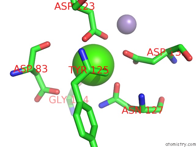
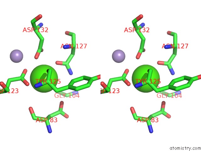
Calcium binding site 1 out of 4 in 1rir
Go back to
Calcium binding site 1 out
of 4 in the Crystal Structure of Meso-Tetrasulphonatophenylporphyrin in Complex with Peanut Lectin.
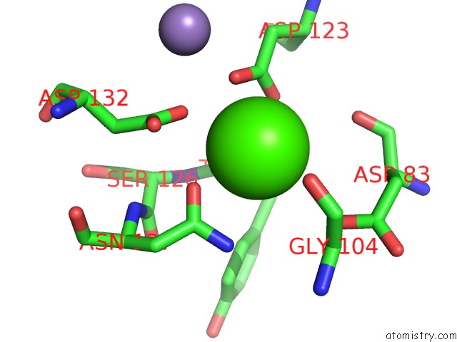
Mono view
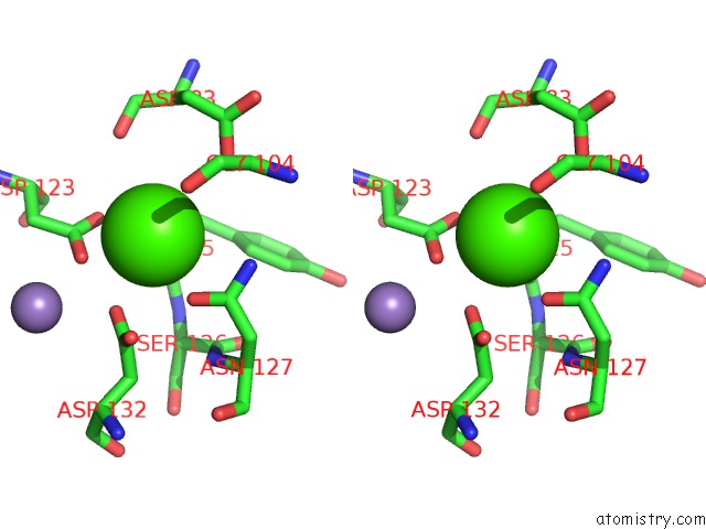
Stereo pair view
Mono view
Stereo pair view
A full contact list of Calcium with other atoms in the Ca binding
site number 1 of Crystal Structure of Meso-Tetrasulphonatophenylporphyrin in Complex with Peanut Lectin. within 5.0Å range:
|
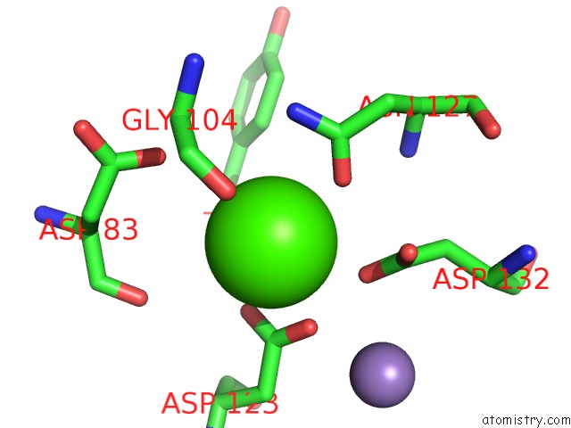
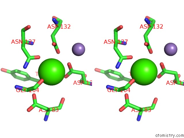
Calcium binding site 2 out of 4 in 1rir
Go back to
Calcium binding site 2 out
of 4 in the Crystal Structure of Meso-Tetrasulphonatophenylporphyrin in Complex with Peanut Lectin.
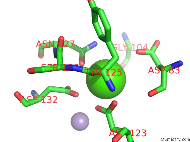
Mono view
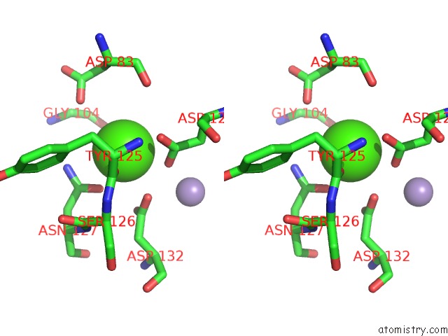
Stereo pair view
Mono view
Stereo pair view
A full contact list of Calcium with other atoms in the Ca binding
site number 2 of Crystal Structure of Meso-Tetrasulphonatophenylporphyrin in Complex with Peanut Lectin. within 5.0Å range:
|
Calcium binding site 3 out of 4 in 1rir
Go back to
Calcium binding site 3 out
of 4 in the Crystal Structure of Meso-Tetrasulphonatophenylporphyrin in Complex with Peanut Lectin.
Mono view
Stereo pair view
Mono view
Stereo pair view
A full contact list of Calcium with other atoms in the Ca binding
site number 3 of Crystal Structure of Meso-Tetrasulphonatophenylporphyrin in Complex with Peanut Lectin. within 5.0Å range:
|
Calcium binding site 4 out of 4 in 1rir
Go back to
Calcium binding site 4 out
of 4 in the Crystal Structure of Meso-Tetrasulphonatophenylporphyrin in Complex with Peanut Lectin.
Mono view
Stereo pair view
Mono view
Stereo pair view
A full contact list of Calcium with other atoms in the Ca binding
site number 4 of Crystal Structure of Meso-Tetrasulphonatophenylporphyrin in Complex with Peanut Lectin. within 5.0Å range:
|
Reference:
M.Goel,
R.S.Damai,
D.K.Sethi,
K.J.Kaur,
B.G.Maiya,
M.J.Swamy,
D.M.Salunke.
Crystal Structures of the Pna-Porphyrin Complex in the Presence and Absence of Lactose: Mapping the Conformational Changes on Lactose Binding, Interacting Surfaces, and Supramolecular Aggregations. Biochemistry V. 44 5588 2005.
ISSN: ISSN 0006-2960
PubMed: 15823017
DOI: 10.1021/BI047377S
Page generated: Thu Jul 11 22:12:21 2024
ISSN: ISSN 0006-2960
PubMed: 15823017
DOI: 10.1021/BI047377S
Last articles
Zn in 9J0NZn in 9J0O
Zn in 9J0P
Zn in 9FJX
Zn in 9EKB
Zn in 9C0F
Zn in 9CAH
Zn in 9CH0
Zn in 9CH3
Zn in 9CH1