Calcium »
PDB 1rpz-1s26 »
1rz5 »
Calcium in PDB 1rz5: Di-Haem Cytochrome C Peroxidase, Form Out
Enzymatic activity of Di-Haem Cytochrome C Peroxidase, Form Out
All present enzymatic activity of Di-Haem Cytochrome C Peroxidase, Form Out:
1.11.1.5;
1.11.1.5;
Protein crystallography data
The structure of Di-Haem Cytochrome C Peroxidase, Form Out, PDB code: 1rz5
was solved by
J.M.Dias,
T.Alves,
C.Bonifacio,
A.S.Pereira,
D.Bourgeois,
I.Moura,
M.J.Romao,
with X-Ray Crystallography technique. A brief refinement statistics is given in the table below:
| Resolution Low / High (Å) | 30.00 / 2.40 |
| Space group | P 64 2 2 |
| Cell size a, b, c (Å), α, β, γ (°) | 150.553, 150.553, 155.349, 90.00, 90.00, 120.00 |
| R / Rfree (%) | 18.6 / 20.9 |
Other elements in 1rz5:
The structure of Di-Haem Cytochrome C Peroxidase, Form Out also contains other interesting chemical elements:
| Iron | (Fe) | 2 atoms |
Calcium Binding Sites:
The binding sites of Calcium atom in the Di-Haem Cytochrome C Peroxidase, Form Out
(pdb code 1rz5). This binding sites where shown within
5.0 Angstroms radius around Calcium atom.
In total only one binding site of Calcium was determined in the Di-Haem Cytochrome C Peroxidase, Form Out, PDB code: 1rz5:
In total only one binding site of Calcium was determined in the Di-Haem Cytochrome C Peroxidase, Form Out, PDB code: 1rz5:
Calcium binding site 1 out of 1 in 1rz5
Go back to
Calcium binding site 1 out
of 1 in the Di-Haem Cytochrome C Peroxidase, Form Out
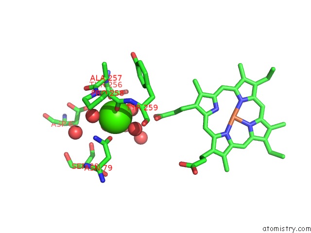
Mono view
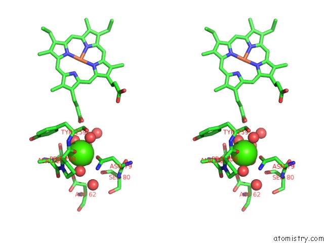
Stereo pair view
Mono view
Stereo pair view
A full contact list of Calcium with other atoms in the Ca binding
site number 1 of Di-Haem Cytochrome C Peroxidase, Form Out within 5.0Å range:
|
Reference:
J.M.Dias,
T.Alves,
C.Bonifacio,
A.S.Pereira,
J.Trincao,
D.Bourgeois,
I.Moura,
M.J.Romao.
Structural Basis For the Mechanism of Ca(2+) Activation of the Di-Heme Cytochrome C Peroxidase From Pseudomonas Nautica 617. Structure V. 12 961 2004.
ISSN: ISSN 0969-2126
PubMed: 15274917
DOI: 10.1016/J.STR.2004.03.025
Page generated: Thu Jul 11 22:20:14 2024
ISSN: ISSN 0969-2126
PubMed: 15274917
DOI: 10.1016/J.STR.2004.03.025
Last articles
Zn in 9J0NZn in 9J0O
Zn in 9J0P
Zn in 9FJX
Zn in 9EKB
Zn in 9C0F
Zn in 9CAH
Zn in 9CH0
Zn in 9CH3
Zn in 9CH1