Calcium »
PDB 1s2n-1scn »
1s6b »
Calcium in PDB 1s6b: X-Ray Crystal Structure of A Complex Formed Between Two Homologous Isoforms of Phospholipase A2 From Naja Naja Sagittifera: Principle of Molecular Association and Inactivation
Enzymatic activity of X-Ray Crystal Structure of A Complex Formed Between Two Homologous Isoforms of Phospholipase A2 From Naja Naja Sagittifera: Principle of Molecular Association and Inactivation
All present enzymatic activity of X-Ray Crystal Structure of A Complex Formed Between Two Homologous Isoforms of Phospholipase A2 From Naja Naja Sagittifera: Principle of Molecular Association and Inactivation:
3.1.1.4;
3.1.1.4;
Protein crystallography data
The structure of X-Ray Crystal Structure of A Complex Formed Between Two Homologous Isoforms of Phospholipase A2 From Naja Naja Sagittifera: Principle of Molecular Association and Inactivation, PDB code: 1s6b
was solved by
T.Jabeen,
S.Sharma,
R.K.Singh,
P.Kaur,
T.P.Singh,
with X-Ray Crystallography technique. A brief refinement statistics is given in the table below:
| Resolution Low / High (Å) | 20.00 / 1.60 |
| Space group | P 41 |
| Cell size a, b, c (Å), α, β, γ (°) | 64.537, 64.537, 57.117, 90.00, 90.00, 90.00 |
| R / Rfree (%) | 20.6 / 23.6 |
Calcium Binding Sites:
The binding sites of Calcium atom in the X-Ray Crystal Structure of A Complex Formed Between Two Homologous Isoforms of Phospholipase A2 From Naja Naja Sagittifera: Principle of Molecular Association and Inactivation
(pdb code 1s6b). This binding sites where shown within
5.0 Angstroms radius around Calcium atom.
In total 2 binding sites of Calcium where determined in the X-Ray Crystal Structure of A Complex Formed Between Two Homologous Isoforms of Phospholipase A2 From Naja Naja Sagittifera: Principle of Molecular Association and Inactivation, PDB code: 1s6b:
Jump to Calcium binding site number: 1; 2;
In total 2 binding sites of Calcium where determined in the X-Ray Crystal Structure of A Complex Formed Between Two Homologous Isoforms of Phospholipase A2 From Naja Naja Sagittifera: Principle of Molecular Association and Inactivation, PDB code: 1s6b:
Jump to Calcium binding site number: 1; 2;
Calcium binding site 1 out of 2 in 1s6b
Go back to
Calcium binding site 1 out
of 2 in the X-Ray Crystal Structure of A Complex Formed Between Two Homologous Isoforms of Phospholipase A2 From Naja Naja Sagittifera: Principle of Molecular Association and Inactivation
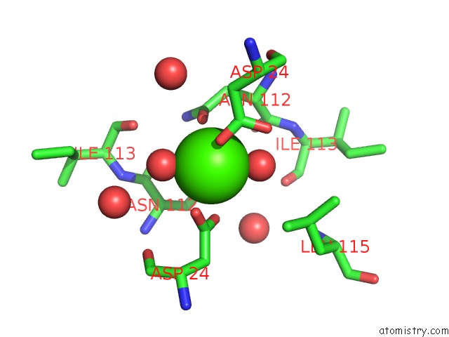
Mono view
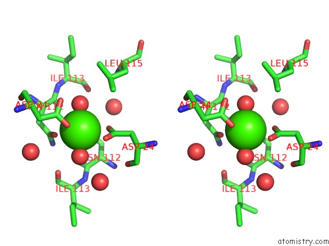
Stereo pair view
Mono view
Stereo pair view
A full contact list of Calcium with other atoms in the Ca binding
site number 1 of X-Ray Crystal Structure of A Complex Formed Between Two Homologous Isoforms of Phospholipase A2 From Naja Naja Sagittifera: Principle of Molecular Association and Inactivation within 5.0Å range:
|
Calcium binding site 2 out of 2 in 1s6b
Go back to
Calcium binding site 2 out
of 2 in the X-Ray Crystal Structure of A Complex Formed Between Two Homologous Isoforms of Phospholipase A2 From Naja Naja Sagittifera: Principle of Molecular Association and Inactivation
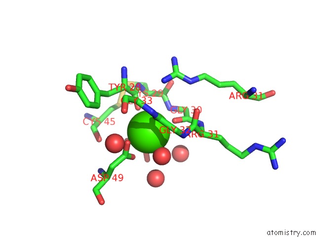
Mono view
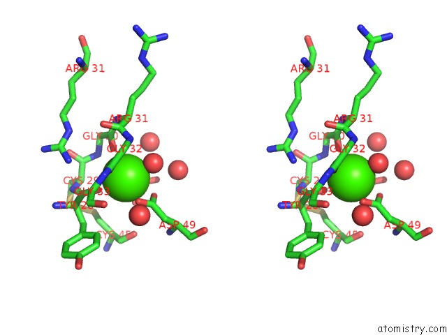
Stereo pair view
Mono view
Stereo pair view
A full contact list of Calcium with other atoms in the Ca binding
site number 2 of X-Ray Crystal Structure of A Complex Formed Between Two Homologous Isoforms of Phospholipase A2 From Naja Naja Sagittifera: Principle of Molecular Association and Inactivation within 5.0Å range:
|
Reference:
T.Jabeen,
S.Sharma,
N.Singh,
R.K.Singh,
P.Kaur,
M.Perbandt,
C.H.Betzel,
A.Srinivasan,
T.P.Singh.
Crystal Structure of A Calcium-Induced Dimer of Two Isoforms of Cobra Phospholipase A2 at 1.6 A Resolution. Proteins V. 59 856 2005.
ISSN: ISSN 0887-3585
PubMed: 15828003
DOI: 10.1002/PROT.20464
Page generated: Thu Jul 11 22:26:56 2024
ISSN: ISSN 0887-3585
PubMed: 15828003
DOI: 10.1002/PROT.20464
Last articles
Zn in 9J0NZn in 9J0O
Zn in 9J0P
Zn in 9FJX
Zn in 9EKB
Zn in 9C0F
Zn in 9CAH
Zn in 9CH0
Zn in 9CH3
Zn in 9CH1