Calcium »
PDB 1scr-1spj »
1sfy »
Calcium in PDB 1sfy: Crystal Structure of Recombinant Erythrina Corallodandron Lectin
Protein crystallography data
The structure of Crystal Structure of Recombinant Erythrina Corallodandron Lectin, PDB code: 1sfy
was solved by
K.A.Kulkarni,
A.Srivastava,
N.Mitra,
A.Surolia,
M.Vijayan,
K.Suguna,
with X-Ray Crystallography technique. A brief refinement statistics is given in the table below:
| Resolution Low / High (Å) | 17.66 / 2.55 |
| Space group | C 1 2 1 |
| Cell size a, b, c (Å), α, β, γ (°) | 87.240, 144.890, 127.660, 90.00, 93.29, 90.00 |
| R / Rfree (%) | 18 / 20.8 |
Other elements in 1sfy:
The structure of Crystal Structure of Recombinant Erythrina Corallodandron Lectin also contains other interesting chemical elements:
| Manganese | (Mn) | 6 atoms |
Calcium Binding Sites:
The binding sites of Calcium atom in the Crystal Structure of Recombinant Erythrina Corallodandron Lectin
(pdb code 1sfy). This binding sites where shown within
5.0 Angstroms radius around Calcium atom.
In total 6 binding sites of Calcium where determined in the Crystal Structure of Recombinant Erythrina Corallodandron Lectin, PDB code: 1sfy:
Jump to Calcium binding site number: 1; 2; 3; 4; 5; 6;
In total 6 binding sites of Calcium where determined in the Crystal Structure of Recombinant Erythrina Corallodandron Lectin, PDB code: 1sfy:
Jump to Calcium binding site number: 1; 2; 3; 4; 5; 6;
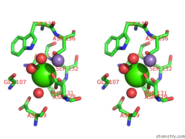
Calcium binding site 1 out of 6 in 1sfy
Go back to
Calcium binding site 1 out
of 6 in the Crystal Structure of Recombinant Erythrina Corallodandron Lectin
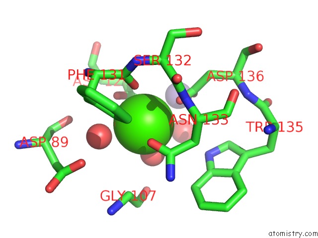
Mono view

Stereo pair view
Mono view
Stereo pair view
A full contact list of Calcium with other atoms in the Ca binding
site number 1 of Crystal Structure of Recombinant Erythrina Corallodandron Lectin within 5.0Å range:
|
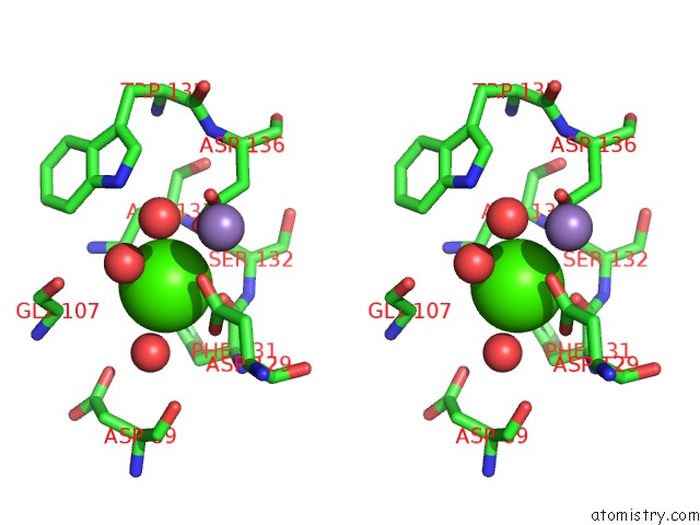
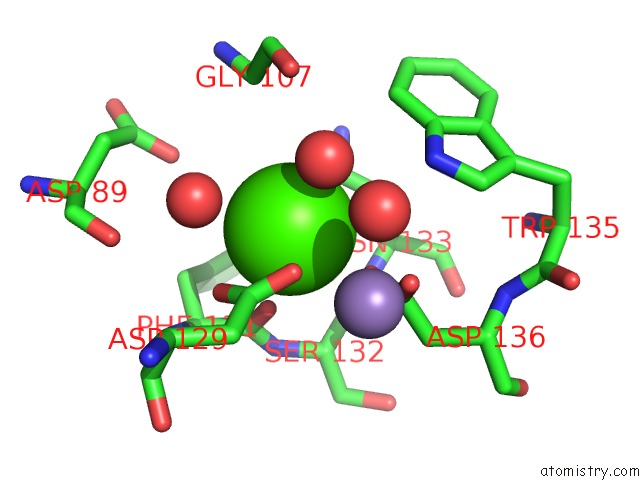
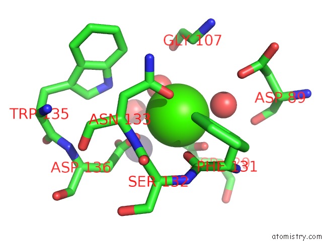
Calcium binding site 2 out of 6 in 1sfy
Go back to
Calcium binding site 2 out
of 6 in the Crystal Structure of Recombinant Erythrina Corallodandron Lectin
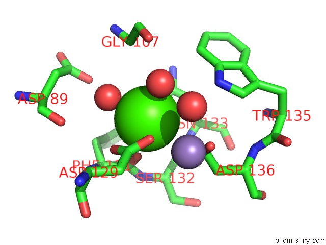
Mono view

Stereo pair view
Mono view
Stereo pair view
A full contact list of Calcium with other atoms in the Ca binding
site number 2 of Crystal Structure of Recombinant Erythrina Corallodandron Lectin within 5.0Å range:
|
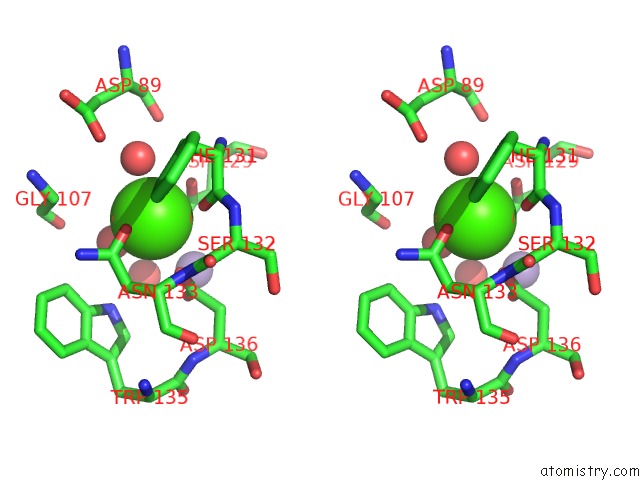
Calcium binding site 3 out of 6 in 1sfy
Go back to
Calcium binding site 3 out
of 6 in the Crystal Structure of Recombinant Erythrina Corallodandron Lectin

Mono view
Stereo pair view
Mono view
Stereo pair view
A full contact list of Calcium with other atoms in the Ca binding
site number 3 of Crystal Structure of Recombinant Erythrina Corallodandron Lectin within 5.0Å range:
|
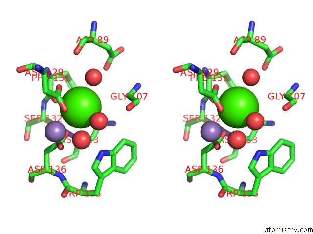
Calcium binding site 4 out of 6 in 1sfy
Go back to
Calcium binding site 4 out
of 6 in the Crystal Structure of Recombinant Erythrina Corallodandron Lectin
Mono view
Stereo pair view
Mono view
Stereo pair view
A full contact list of Calcium with other atoms in the Ca binding
site number 4 of Crystal Structure of Recombinant Erythrina Corallodandron Lectin within 5.0Å range:
|
Calcium binding site 5 out of 6 in 1sfy
Go back to
Calcium binding site 5 out
of 6 in the Crystal Structure of Recombinant Erythrina Corallodandron Lectin
Mono view
Stereo pair view
Mono view
Stereo pair view
A full contact list of Calcium with other atoms in the Ca binding
site number 5 of Crystal Structure of Recombinant Erythrina Corallodandron Lectin within 5.0Å range:
|
Calcium binding site 6 out of 6 in 1sfy
Go back to
Calcium binding site 6 out
of 6 in the Crystal Structure of Recombinant Erythrina Corallodandron Lectin

Mono view
Stereo pair view
Mono view
Stereo pair view
A full contact list of Calcium with other atoms in the Ca binding
site number 6 of Crystal Structure of Recombinant Erythrina Corallodandron Lectin within 5.0Å range:
|
Reference:
K.A.Kulkarni,
A.Srivastava,
N.Mitra,
N.Sharon,
A.Surolia,
M.Vijayan,
K.Suguna.
Effect of Glycosylation on the Structure of Erythrina Corallodendron Lectin. Proteins V. 56 821 2004.
ISSN: ISSN 0887-3585
PubMed: 15281133
DOI: 10.1002/PROT.20168
Page generated: Thu Jul 11 22:37:05 2024
ISSN: ISSN 0887-3585
PubMed: 15281133
DOI: 10.1002/PROT.20168
Last articles
Zn in 9J0NZn in 9J0O
Zn in 9J0P
Zn in 9FJX
Zn in 9EKB
Zn in 9C0F
Zn in 9CAH
Zn in 9CH0
Zn in 9CH3
Zn in 9CH1