Calcium »
PDB 1scr-1spj »
1sl6 »
Calcium in PDB 1sl6: Crystal Structure of A Fragment of Dc-Signr (Containg the Carbohydrate Recognition Domain and Two Repeats of the Neck) Complexed with Lewis-X.
Protein crystallography data
The structure of Crystal Structure of A Fragment of Dc-Signr (Containg the Carbohydrate Recognition Domain and Two Repeats of the Neck) Complexed with Lewis-X., PDB code: 1sl6
was solved by
Y.Guo,
H.Feinberg,
E.Conroy,
D.A.Mitchell,
R.Alvarez,
O.Blixt,
M.E.Taylor,
W.I.Weis,
K.Drickamer,
with X-Ray Crystallography technique. A brief refinement statistics is given in the table below:
| Resolution Low / High (Å) | 41.96 / 2.25 |
| Space group | P 32 2 1 |
| Cell size a, b, c (Å), α, β, γ (°) | 153.750, 153.750, 128.702, 90.00, 90.00, 120.00 |
| R / Rfree (%) | 21.9 / 25.6 |
Calcium Binding Sites:
Pages:
>>> Page 1 <<< Page 2, Binding sites: 11 - 20; Page 3, Binding sites: 21 - 24;Binding sites:
The binding sites of Calcium atom in the Crystal Structure of A Fragment of Dc-Signr (Containg the Carbohydrate Recognition Domain and Two Repeats of the Neck) Complexed with Lewis-X. (pdb code 1sl6). This binding sites where shown within 5.0 Angstroms radius around Calcium atom.In total 24 binding sites of Calcium where determined in the Crystal Structure of A Fragment of Dc-Signr (Containg the Carbohydrate Recognition Domain and Two Repeats of the Neck) Complexed with Lewis-X., PDB code: 1sl6:
Jump to Calcium binding site number: 1; 2; 3; 4; 5; 6; 7; 8; 9; 10;
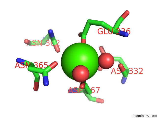
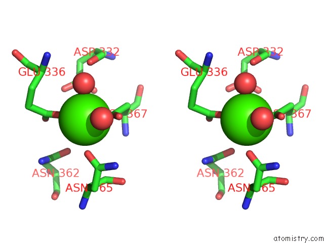
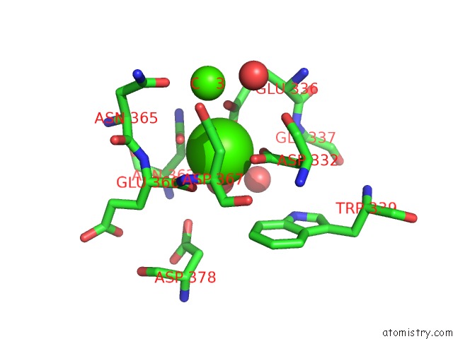
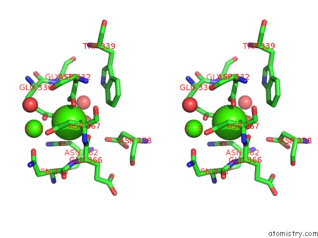
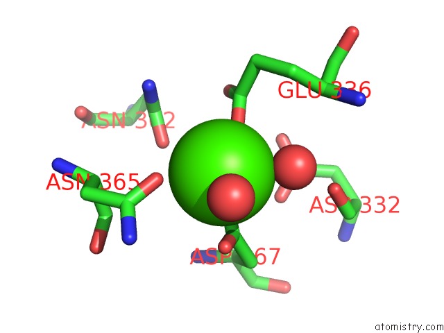
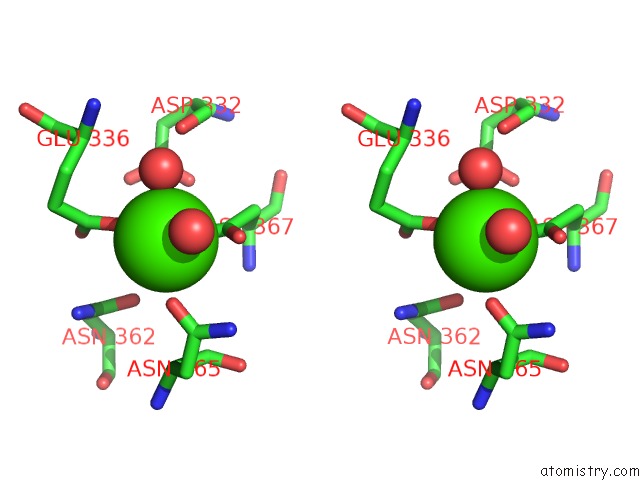
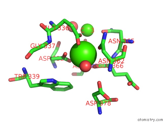
Calcium binding site 1 out of 24 in 1sl6
Go back to
Calcium binding site 1 out
of 24 in the Crystal Structure of A Fragment of Dc-Signr (Containg the Carbohydrate Recognition Domain and Two Repeats of the Neck) Complexed with Lewis-X.
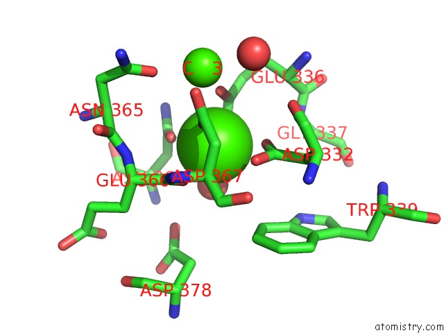
Mono view
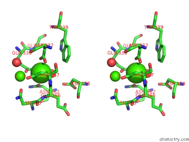
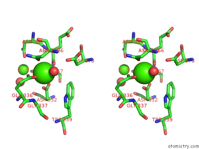
Stereo pair view
Mono view
Stereo pair view
A full contact list of Calcium with other atoms in the Ca binding
site number 1 of Crystal Structure of A Fragment of Dc-Signr (Containg the Carbohydrate Recognition Domain and Two Repeats of the Neck) Complexed with Lewis-X. within 5.0Å range:
|
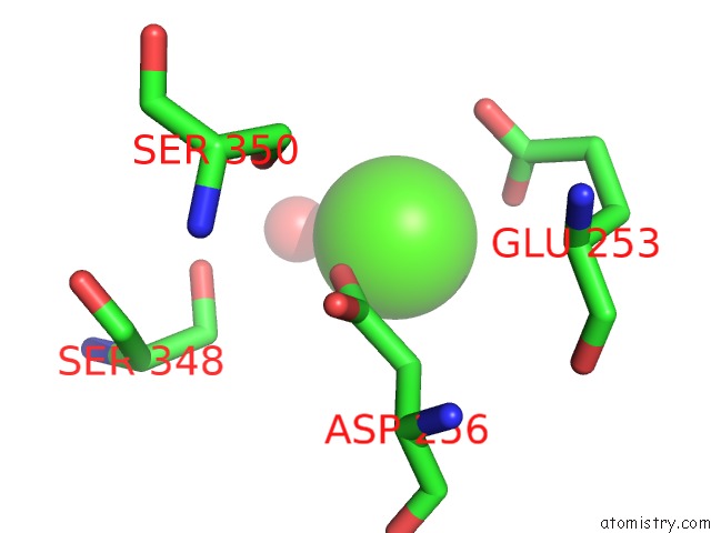
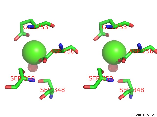
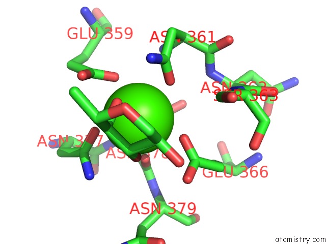
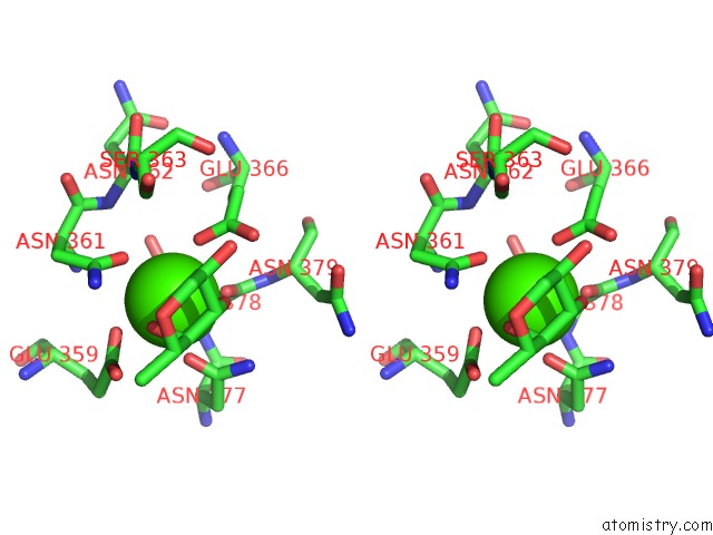
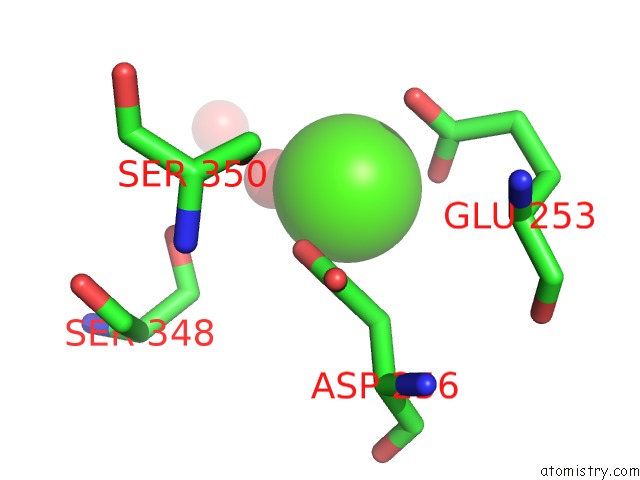
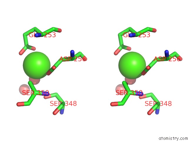
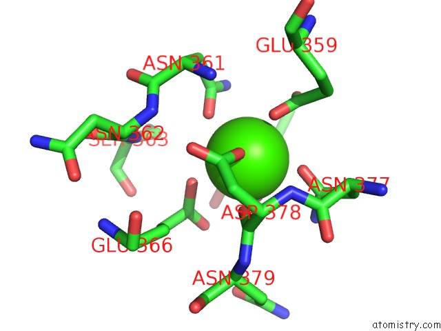
Calcium binding site 2 out of 24 in 1sl6
Go back to
Calcium binding site 2 out
of 24 in the Crystal Structure of A Fragment of Dc-Signr (Containg the Carbohydrate Recognition Domain and Two Repeats of the Neck) Complexed with Lewis-X.
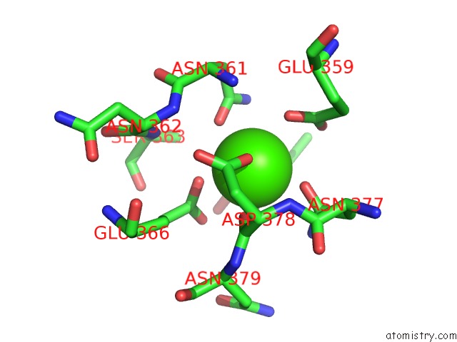
Mono view
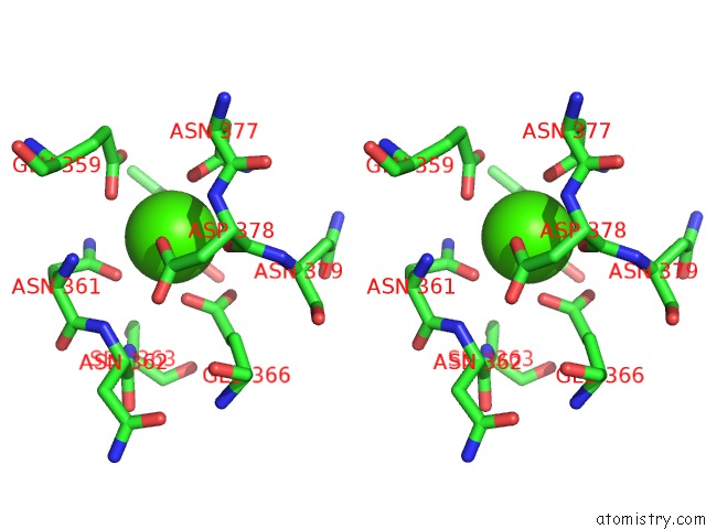
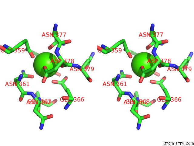
Stereo pair view
Mono view
Stereo pair view
A full contact list of Calcium with other atoms in the Ca binding
site number 2 of Crystal Structure of A Fragment of Dc-Signr (Containg the Carbohydrate Recognition Domain and Two Repeats of the Neck) Complexed with Lewis-X. within 5.0Å range:
|
Calcium binding site 3 out of 24 in 1sl6
Go back to
Calcium binding site 3 out
of 24 in the Crystal Structure of A Fragment of Dc-Signr (Containg the Carbohydrate Recognition Domain and Two Repeats of the Neck) Complexed with Lewis-X.
Mono view
Stereo pair view
Mono view
Stereo pair view
A full contact list of Calcium with other atoms in the Ca binding
site number 3 of Crystal Structure of A Fragment of Dc-Signr (Containg the Carbohydrate Recognition Domain and Two Repeats of the Neck) Complexed with Lewis-X. within 5.0Å range:
|
Calcium binding site 4 out of 24 in 1sl6
Go back to
Calcium binding site 4 out
of 24 in the Crystal Structure of A Fragment of Dc-Signr (Containg the Carbohydrate Recognition Domain and Two Repeats of the Neck) Complexed with Lewis-X.
Mono view
Stereo pair view
Mono view
Stereo pair view
A full contact list of Calcium with other atoms in the Ca binding
site number 4 of Crystal Structure of A Fragment of Dc-Signr (Containg the Carbohydrate Recognition Domain and Two Repeats of the Neck) Complexed with Lewis-X. within 5.0Å range:
|
Calcium binding site 5 out of 24 in 1sl6
Go back to
Calcium binding site 5 out
of 24 in the Crystal Structure of A Fragment of Dc-Signr (Containg the Carbohydrate Recognition Domain and Two Repeats of the Neck) Complexed with Lewis-X.
Mono view
Stereo pair view
Mono view
Stereo pair view
A full contact list of Calcium with other atoms in the Ca binding
site number 5 of Crystal Structure of A Fragment of Dc-Signr (Containg the Carbohydrate Recognition Domain and Two Repeats of the Neck) Complexed with Lewis-X. within 5.0Å range:
|
Calcium binding site 6 out of 24 in 1sl6
Go back to
Calcium binding site 6 out
of 24 in the Crystal Structure of A Fragment of Dc-Signr (Containg the Carbohydrate Recognition Domain and Two Repeats of the Neck) Complexed with Lewis-X.
Mono view
Stereo pair view
Mono view
Stereo pair view
A full contact list of Calcium with other atoms in the Ca binding
site number 6 of Crystal Structure of A Fragment of Dc-Signr (Containg the Carbohydrate Recognition Domain and Two Repeats of the Neck) Complexed with Lewis-X. within 5.0Å range:
|
Calcium binding site 7 out of 24 in 1sl6
Go back to
Calcium binding site 7 out
of 24 in the Crystal Structure of A Fragment of Dc-Signr (Containg the Carbohydrate Recognition Domain and Two Repeats of the Neck) Complexed with Lewis-X.
Mono view
Stereo pair view
Mono view
Stereo pair view
A full contact list of Calcium with other atoms in the Ca binding
site number 7 of Crystal Structure of A Fragment of Dc-Signr (Containg the Carbohydrate Recognition Domain and Two Repeats of the Neck) Complexed with Lewis-X. within 5.0Å range:
|
Calcium binding site 8 out of 24 in 1sl6
Go back to
Calcium binding site 8 out
of 24 in the Crystal Structure of A Fragment of Dc-Signr (Containg the Carbohydrate Recognition Domain and Two Repeats of the Neck) Complexed with Lewis-X.
Mono view
Stereo pair view
Mono view
Stereo pair view
A full contact list of Calcium with other atoms in the Ca binding
site number 8 of Crystal Structure of A Fragment of Dc-Signr (Containg the Carbohydrate Recognition Domain and Two Repeats of the Neck) Complexed with Lewis-X. within 5.0Å range:
|
Calcium binding site 9 out of 24 in 1sl6
Go back to
Calcium binding site 9 out
of 24 in the Crystal Structure of A Fragment of Dc-Signr (Containg the Carbohydrate Recognition Domain and Two Repeats of the Neck) Complexed with Lewis-X.
Mono view
Stereo pair view
Mono view
Stereo pair view
A full contact list of Calcium with other atoms in the Ca binding
site number 9 of Crystal Structure of A Fragment of Dc-Signr (Containg the Carbohydrate Recognition Domain and Two Repeats of the Neck) Complexed with Lewis-X. within 5.0Å range:
|
Calcium binding site 10 out of 24 in 1sl6
Go back to
Calcium binding site 10 out
of 24 in the Crystal Structure of A Fragment of Dc-Signr (Containg the Carbohydrate Recognition Domain and Two Repeats of the Neck) Complexed with Lewis-X.
Mono view
Stereo pair view
Mono view
Stereo pair view
A full contact list of Calcium with other atoms in the Ca binding
site number 10 of Crystal Structure of A Fragment of Dc-Signr (Containg the Carbohydrate Recognition Domain and Two Repeats of the Neck) Complexed with Lewis-X. within 5.0Å range:
|
Reference:
Y.Guo,
H.Feinberg,
E.Conroy,
D.A.Mitchell,
R.Alvarez,
O.Blixt,
M.E.Taylor,
W.I.Weis,
K.Drickamer.
Structural Basis For Distinct Ligand-Binding and Targeting Properties of the Receptors Dc-Sign and Dc-Signr Nat.Struct.Mol.Biol. V. 11 591 2004.
ISSN: ISSN 1545-9993
PubMed: 15195147
DOI: 10.1038/NSMB784
Page generated: Tue Jul 8 01:59:30 2025
ISSN: ISSN 1545-9993
PubMed: 15195147
DOI: 10.1038/NSMB784
Last articles
Cl in 5KA7Cl in 5KA2
Cl in 5KA3
Cl in 5KA0
Cl in 5K9V
Cl in 5K9W
Cl in 5KA1
Cl in 5K9D
Cl in 5K9C
Cl in 5K92