Calcium »
PDB 1scr-1spj »
1sln »
Calcium in PDB 1sln: Crystal Structure of the Catalytic Domain of Human Fibroblast Stromelysin-1 Inhibited with the N-Carboxy-Alkyl Inhibitor L-702,842
Enzymatic activity of Crystal Structure of the Catalytic Domain of Human Fibroblast Stromelysin-1 Inhibited with the N-Carboxy-Alkyl Inhibitor L-702,842
All present enzymatic activity of Crystal Structure of the Catalytic Domain of Human Fibroblast Stromelysin-1 Inhibited with the N-Carboxy-Alkyl Inhibitor L-702,842:
3.4.24.17;
3.4.24.17;
Protein crystallography data
The structure of Crystal Structure of the Catalytic Domain of Human Fibroblast Stromelysin-1 Inhibited with the N-Carboxy-Alkyl Inhibitor L-702,842, PDB code: 1sln
was solved by
J.W.Becker,
with X-Ray Crystallography technique. A brief refinement statistics is given in the table below:
| Resolution Low / High (Å) | 8.00 / 2.27 |
| Space group | P 31 2 1 |
| Cell size a, b, c (Å), α, β, γ (°) | 47.230, 47.230, 150.850, 90.00, 90.00, 120.00 |
| R / Rfree (%) | 22.6 / 29.9 |
Other elements in 1sln:
The structure of Crystal Structure of the Catalytic Domain of Human Fibroblast Stromelysin-1 Inhibited with the N-Carboxy-Alkyl Inhibitor L-702,842 also contains other interesting chemical elements:
| Zinc | (Zn) | 2 atoms |
Calcium Binding Sites:
The binding sites of Calcium atom in the Crystal Structure of the Catalytic Domain of Human Fibroblast Stromelysin-1 Inhibited with the N-Carboxy-Alkyl Inhibitor L-702,842
(pdb code 1sln). This binding sites where shown within
5.0 Angstroms radius around Calcium atom.
In total 3 binding sites of Calcium where determined in the Crystal Structure of the Catalytic Domain of Human Fibroblast Stromelysin-1 Inhibited with the N-Carboxy-Alkyl Inhibitor L-702,842, PDB code: 1sln:
Jump to Calcium binding site number: 1; 2; 3;
In total 3 binding sites of Calcium where determined in the Crystal Structure of the Catalytic Domain of Human Fibroblast Stromelysin-1 Inhibited with the N-Carboxy-Alkyl Inhibitor L-702,842, PDB code: 1sln:
Jump to Calcium binding site number: 1; 2; 3;
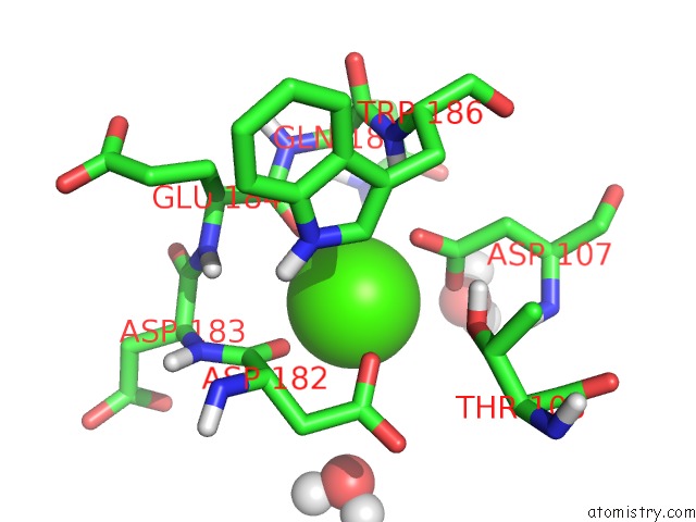
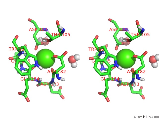
Calcium binding site 1 out of 3 in 1sln
Go back to
Calcium binding site 1 out
of 3 in the Crystal Structure of the Catalytic Domain of Human Fibroblast Stromelysin-1 Inhibited with the N-Carboxy-Alkyl Inhibitor L-702,842
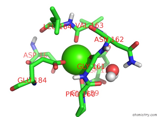
Mono view
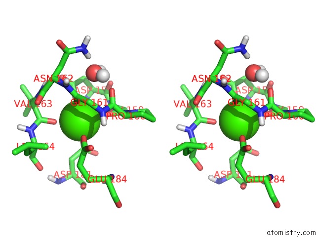
Stereo pair view
Mono view
Stereo pair view
A full contact list of Calcium with other atoms in the Ca binding
site number 1 of Crystal Structure of the Catalytic Domain of Human Fibroblast Stromelysin-1 Inhibited with the N-Carboxy-Alkyl Inhibitor L-702,842 within 5.0Å range:
|
Calcium binding site 2 out of 3 in 1sln
Go back to
Calcium binding site 2 out
of 3 in the Crystal Structure of the Catalytic Domain of Human Fibroblast Stromelysin-1 Inhibited with the N-Carboxy-Alkyl Inhibitor L-702,842
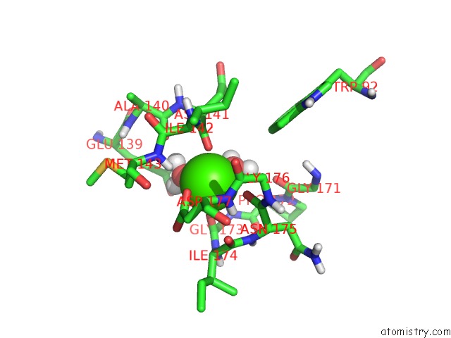
Mono view
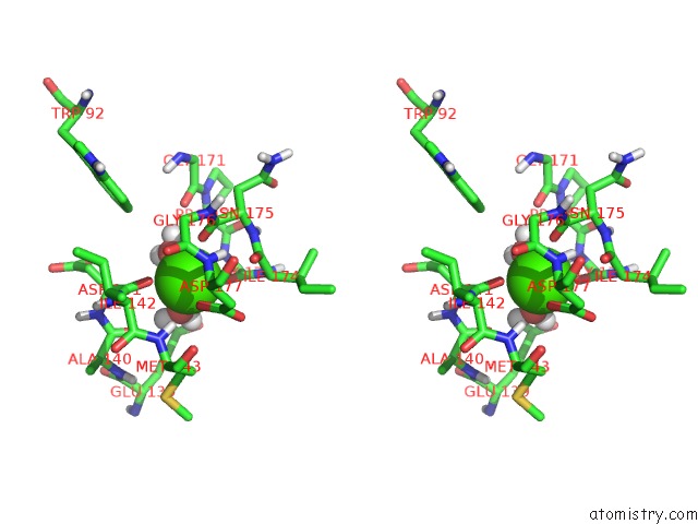
Stereo pair view
Mono view
Stereo pair view
A full contact list of Calcium with other atoms in the Ca binding
site number 2 of Crystal Structure of the Catalytic Domain of Human Fibroblast Stromelysin-1 Inhibited with the N-Carboxy-Alkyl Inhibitor L-702,842 within 5.0Å range:
|
Calcium binding site 3 out of 3 in 1sln
Go back to
Calcium binding site 3 out
of 3 in the Crystal Structure of the Catalytic Domain of Human Fibroblast Stromelysin-1 Inhibited with the N-Carboxy-Alkyl Inhibitor L-702,842
Mono view
Stereo pair view
Mono view
Stereo pair view
A full contact list of Calcium with other atoms in the Ca binding
site number 3 of Crystal Structure of the Catalytic Domain of Human Fibroblast Stromelysin-1 Inhibited with the N-Carboxy-Alkyl Inhibitor L-702,842 within 5.0Å range:
|
Reference:
J.W.Becker,
A.I.Marcy,
L.L.Rokosz,
M.G.Axel,
J.J.Burbaum,
P.M.Fitzgerald,
P.M.Cameron,
C.K.Esser,
W.K.Hagmann,
J.D.Hermes,
J.P.Springer.
Stromelysin-1: Three-Dimensional Structure of the Inhibited Catalytic Domain and of the C-Truncated Proenzyme. Protein Sci. V. 4 1966 1995.
ISSN: ISSN 0961-8368
PubMed: 8535233
Page generated: Tue Jul 8 02:00:25 2025
ISSN: ISSN 0961-8368
PubMed: 8535233
Last articles
Cl in 8CQFCl in 8CPI
Cl in 8CQ3
Cl in 8COV
Cl in 8CPH
Cl in 8CNO
Cl in 8CO7
Cl in 8CNZ
Cl in 8COT
Cl in 8COJ