Calcium »
PDB 1scr-1spj »
1smv »
Calcium in PDB 1smv: Primary Structure of Sesbania Mosaic Virus Coat Protein: Its Implications to the Assembly and Architecture of the Virus
Protein crystallography data
The structure of Primary Structure of Sesbania Mosaic Virus Coat Protein: Its Implications to the Assembly and Architecture of the Virus, PDB code: 1smv
was solved by
M.Bhuvaneshwari,
M.R.N.Murthy,
with X-Ray Crystallography technique. A brief refinement statistics is given in the table below:
| Resolution Low / High (Å) | 10.00 / 3.00 |
| Space group | R 3 1 |
| Cell size a, b, c (Å), α, β, γ (°) | 291.460, 291.460, 291.460, 61.95, 61.95, 61.95 |
| R / Rfree (%) | 22.7 / n/a |
Calcium Binding Sites:
The binding sites of Calcium atom in the Primary Structure of Sesbania Mosaic Virus Coat Protein: Its Implications to the Assembly and Architecture of the Virus
(pdb code 1smv). This binding sites where shown within
5.0 Angstroms radius around Calcium atom.
In total 4 binding sites of Calcium where determined in the Primary Structure of Sesbania Mosaic Virus Coat Protein: Its Implications to the Assembly and Architecture of the Virus, PDB code: 1smv:
Jump to Calcium binding site number: 1; 2; 3; 4;
In total 4 binding sites of Calcium where determined in the Primary Structure of Sesbania Mosaic Virus Coat Protein: Its Implications to the Assembly and Architecture of the Virus, PDB code: 1smv:
Jump to Calcium binding site number: 1; 2; 3; 4;
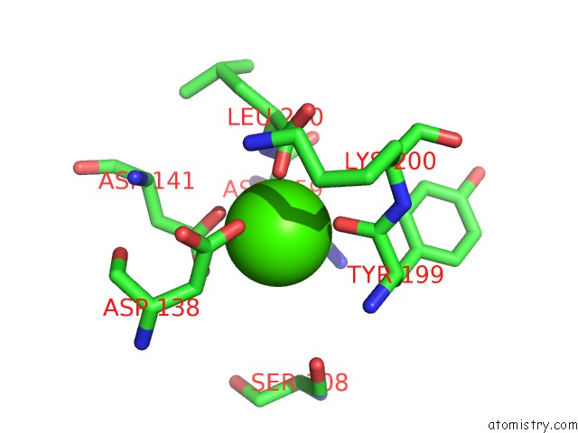
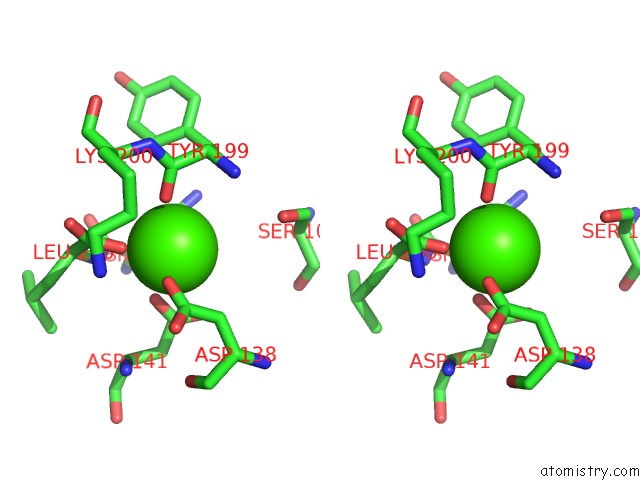
Calcium binding site 1 out of 4 in 1smv
Go back to
Calcium binding site 1 out
of 4 in the Primary Structure of Sesbania Mosaic Virus Coat Protein: Its Implications to the Assembly and Architecture of the Virus
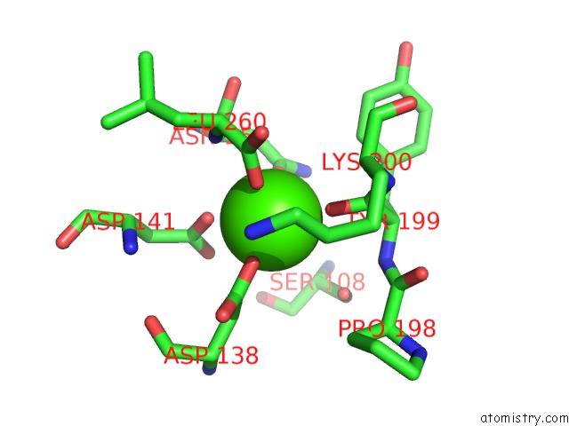
Mono view
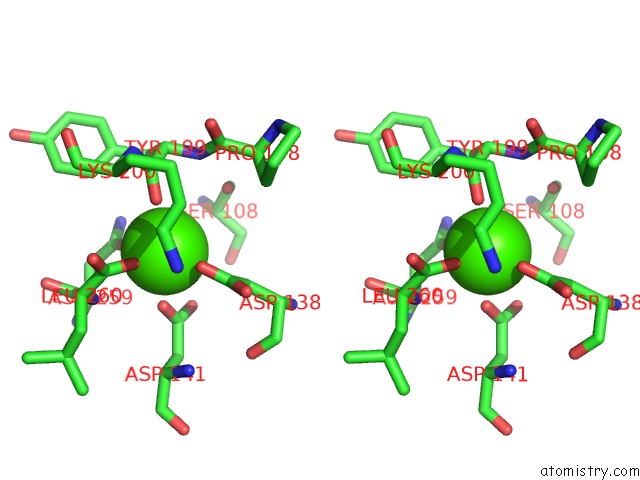
Stereo pair view
Mono view
Stereo pair view
A full contact list of Calcium with other atoms in the Ca binding
site number 1 of Primary Structure of Sesbania Mosaic Virus Coat Protein: Its Implications to the Assembly and Architecture of the Virus within 5.0Å range:
|
Calcium binding site 2 out of 4 in 1smv
Go back to
Calcium binding site 2 out
of 4 in the Primary Structure of Sesbania Mosaic Virus Coat Protein: Its Implications to the Assembly and Architecture of the Virus
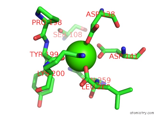
Mono view
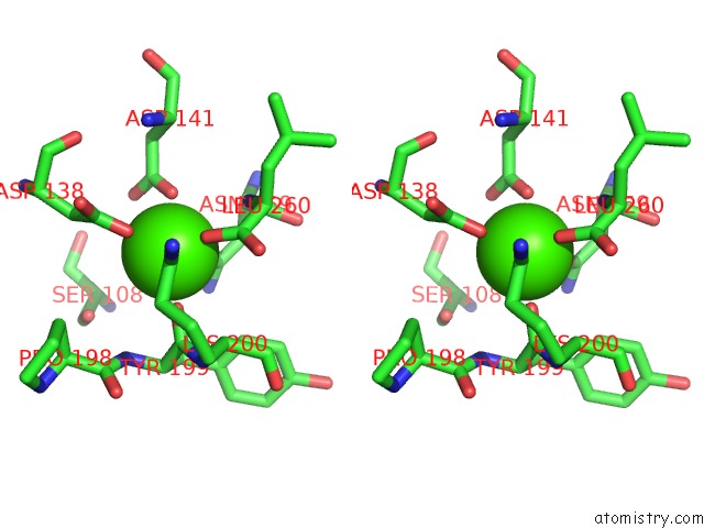
Stereo pair view
Mono view
Stereo pair view
A full contact list of Calcium with other atoms in the Ca binding
site number 2 of Primary Structure of Sesbania Mosaic Virus Coat Protein: Its Implications to the Assembly and Architecture of the Virus within 5.0Å range:
|
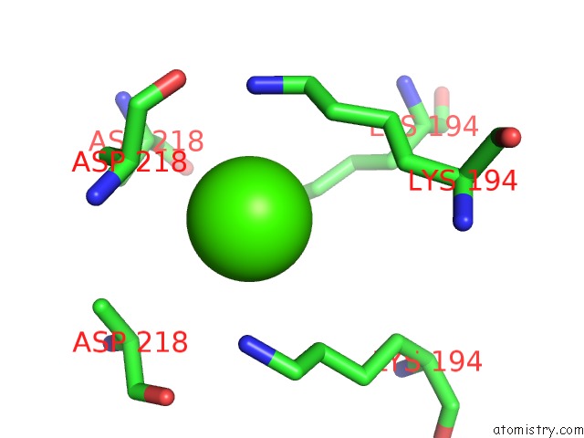
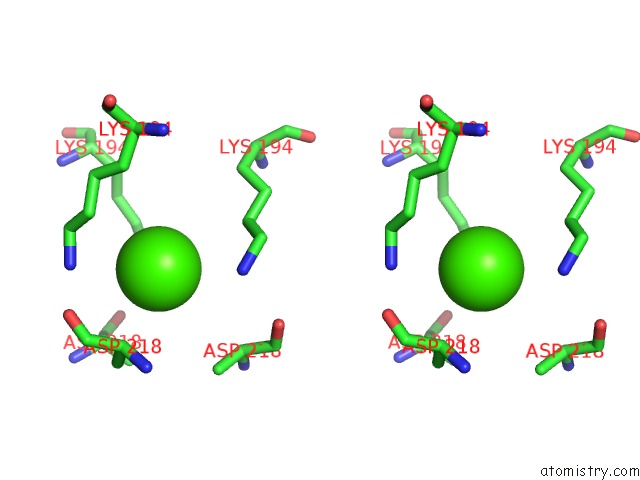
Calcium binding site 3 out of 4 in 1smv
Go back to
Calcium binding site 3 out
of 4 in the Primary Structure of Sesbania Mosaic Virus Coat Protein: Its Implications to the Assembly and Architecture of the Virus
Mono view
Stereo pair view
Mono view
Stereo pair view
A full contact list of Calcium with other atoms in the Ca binding
site number 3 of Primary Structure of Sesbania Mosaic Virus Coat Protein: Its Implications to the Assembly and Architecture of the Virus within 5.0Å range:
|
Calcium binding site 4 out of 4 in 1smv
Go back to
Calcium binding site 4 out
of 4 in the Primary Structure of Sesbania Mosaic Virus Coat Protein: Its Implications to the Assembly and Architecture of the Virus
Mono view
Stereo pair view
Mono view
Stereo pair view
A full contact list of Calcium with other atoms in the Ca binding
site number 4 of Primary Structure of Sesbania Mosaic Virus Coat Protein: Its Implications to the Assembly and Architecture of the Virus within 5.0Å range:
|
Reference:
M.Bhuvaneshwari,
H.S.Subramanya,
K.Gopinath,
H.S.Savithri,
M.V.Nayudu,
M.R.Murthy.
Structure of Sesbania Mosaic Virus at 3 A Resolution. Structure V. 3 1021 1995.
ISSN: ISSN 0969-2126
PubMed: 8589997
DOI: 10.1016/S0969-2126(01)00238-6
Page generated: Tue Jul 8 02:01:42 2025
ISSN: ISSN 0969-2126
PubMed: 8589997
DOI: 10.1016/S0969-2126(01)00238-6
Last articles
Cl in 5KGMCl in 5KGR
Cl in 5KH3
Cl in 5KGT
Cl in 5KGN
Cl in 5KGG
Cl in 5KGI
Cl in 5KGL
Cl in 5KGH
Cl in 5KC9