Calcium »
PDB 1spu-1t5s »
1st2 »
Calcium in PDB 1st2: The Three-Dimensional Structure of Bacillus Amyloliquefaciens Subtilisin at 1.8 Angstroms and An Analysis of the Structural Consequences of Peroxide Inactivation
Enzymatic activity of The Three-Dimensional Structure of Bacillus Amyloliquefaciens Subtilisin at 1.8 Angstroms and An Analysis of the Structural Consequences of Peroxide Inactivation
All present enzymatic activity of The Three-Dimensional Structure of Bacillus Amyloliquefaciens Subtilisin at 1.8 Angstroms and An Analysis of the Structural Consequences of Peroxide Inactivation:
3.4.21.14;
3.4.21.14;
Protein crystallography data
The structure of The Three-Dimensional Structure of Bacillus Amyloliquefaciens Subtilisin at 1.8 Angstroms and An Analysis of the Structural Consequences of Peroxide Inactivation, PDB code: 1st2
was solved by
R.Bott,
with X-Ray Crystallography technique. A brief refinement statistics is given in the table below:
| Resolution Low / High (Å) | 10.00 / 2.00 |
| Space group | P 21 21 21 |
| Cell size a, b, c (Å), α, β, γ (°) | 39.300, 72.800, 75.350, 90.00, 90.00, 90.00 |
| R / Rfree (%) | n/a / n/a |
Calcium Binding Sites:
The binding sites of Calcium atom in the The Three-Dimensional Structure of Bacillus Amyloliquefaciens Subtilisin at 1.8 Angstroms and An Analysis of the Structural Consequences of Peroxide Inactivation
(pdb code 1st2). This binding sites where shown within
5.0 Angstroms radius around Calcium atom.
In total 2 binding sites of Calcium where determined in the The Three-Dimensional Structure of Bacillus Amyloliquefaciens Subtilisin at 1.8 Angstroms and An Analysis of the Structural Consequences of Peroxide Inactivation, PDB code: 1st2:
Jump to Calcium binding site number: 1; 2;
In total 2 binding sites of Calcium where determined in the The Three-Dimensional Structure of Bacillus Amyloliquefaciens Subtilisin at 1.8 Angstroms and An Analysis of the Structural Consequences of Peroxide Inactivation, PDB code: 1st2:
Jump to Calcium binding site number: 1; 2;
Calcium binding site 1 out of 2 in 1st2
Go back to
Calcium binding site 1 out
of 2 in the The Three-Dimensional Structure of Bacillus Amyloliquefaciens Subtilisin at 1.8 Angstroms and An Analysis of the Structural Consequences of Peroxide Inactivation
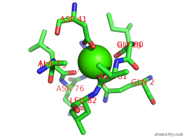
Mono view
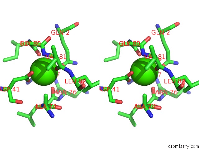
Stereo pair view
Mono view
Stereo pair view
A full contact list of Calcium with other atoms in the Ca binding
site number 1 of The Three-Dimensional Structure of Bacillus Amyloliquefaciens Subtilisin at 1.8 Angstroms and An Analysis of the Structural Consequences of Peroxide Inactivation within 5.0Å range:
|
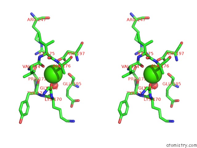
Calcium binding site 2 out of 2 in 1st2
Go back to
Calcium binding site 2 out
of 2 in the The Three-Dimensional Structure of Bacillus Amyloliquefaciens Subtilisin at 1.8 Angstroms and An Analysis of the Structural Consequences of Peroxide Inactivation
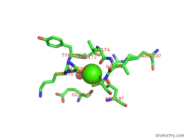
Mono view
Stereo pair view
Mono view
Stereo pair view
A full contact list of Calcium with other atoms in the Ca binding
site number 2 of The Three-Dimensional Structure of Bacillus Amyloliquefaciens Subtilisin at 1.8 Angstroms and An Analysis of the Structural Consequences of Peroxide Inactivation within 5.0Å range:
|
Reference:
R.Bott,
M.Ultsch,
A.Kossiakoff,
T.Graycar,
B.Katz,
S.Power.
The Three-Dimensional Structure of Bacillus Amyloliquefaciens Subtilisin at 1.8 A and An Analysis of the Structural Consequences of Peroxide Inactivation. J.Biol.Chem. V. 263 7895 1988.
ISSN: ISSN 0021-9258
PubMed: 3286644
Page generated: Thu Jul 11 22:48:46 2024
ISSN: ISSN 0021-9258
PubMed: 3286644
Last articles
Zn in 9MJ5Zn in 9HNW
Zn in 9G0L
Zn in 9FNE
Zn in 9DZN
Zn in 9E0I
Zn in 9D32
Zn in 9DAK
Zn in 8ZXC
Zn in 8ZUF