Calcium »
PDB 1spu-1t5s »
1svn »
Calcium in PDB 1svn: Savinase
Enzymatic activity of Savinase
All present enzymatic activity of Savinase:
3.4.21.62;
3.4.21.62;
Protein crystallography data
The structure of Savinase, PDB code: 1svn
was solved by
C.Betzel,
S.Klupsch,
G.Papendorf,
S.Hastrup,
S.Branner,
K.S.Wilson,
with X-Ray Crystallography technique. A brief refinement statistics is given in the table below:
| Resolution Low / High (Å) | 10.00 / 1.40 |
| Space group | P 1 21 1 |
| Cell size a, b, c (Å), α, β, γ (°) | 40.470, 64.240, 42.890, 90.00, 118.80, 90.00 |
| R / Rfree (%) | 17.4 / n/a |
Calcium Binding Sites:
The binding sites of Calcium atom in the Savinase
(pdb code 1svn). This binding sites where shown within
5.0 Angstroms radius around Calcium atom.
In total 2 binding sites of Calcium where determined in the Savinase, PDB code: 1svn:
Jump to Calcium binding site number: 1; 2;
In total 2 binding sites of Calcium where determined in the Savinase, PDB code: 1svn:
Jump to Calcium binding site number: 1; 2;
Calcium binding site 1 out of 2 in 1svn
Go back to
Calcium binding site 1 out
of 2 in the Savinase
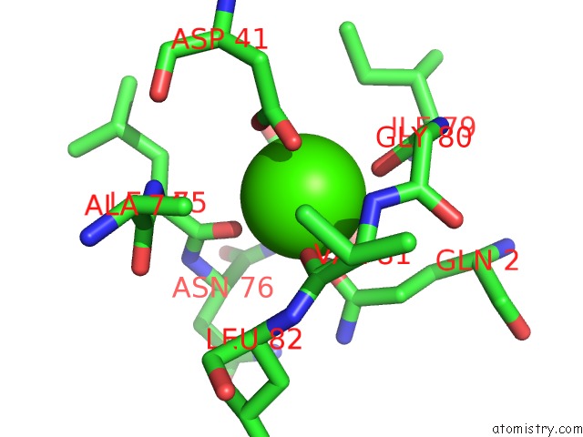
Mono view
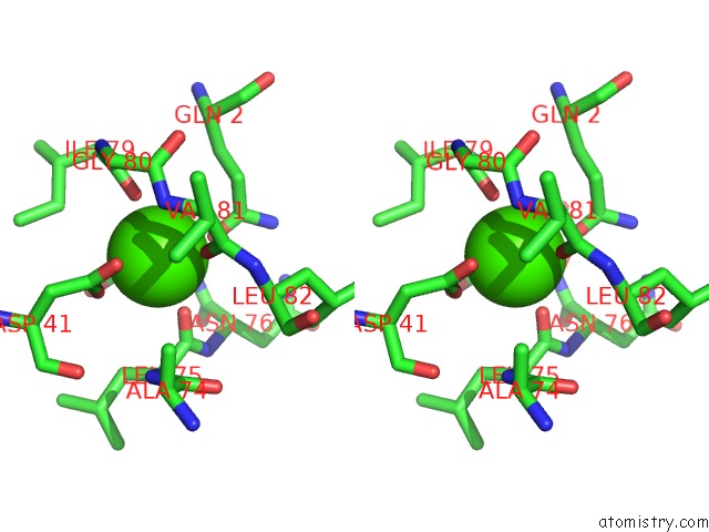
Stereo pair view
Mono view
Stereo pair view
A full contact list of Calcium with other atoms in the Ca binding
site number 1 of Savinase within 5.0Å range:
|
Calcium binding site 2 out of 2 in 1svn
Go back to
Calcium binding site 2 out
of 2 in the Savinase
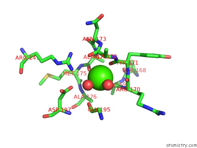
Mono view
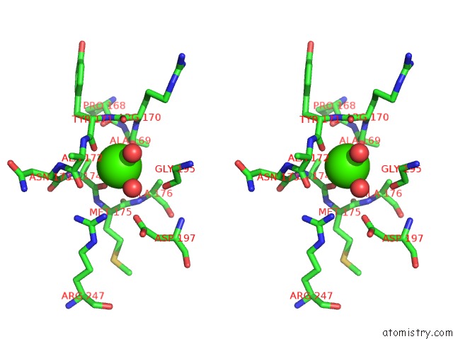
Stereo pair view
Mono view
Stereo pair view
A full contact list of Calcium with other atoms in the Ca binding
site number 2 of Savinase within 5.0Å range:
|
Reference:
C.Betzel,
S.Klupsch,
G.Papendorf,
S.Hastrup,
S.Branner,
K.S.Wilson.
Crystal Structure of the Alkaline Proteinase Savinase From Bacillus Lentus at 1.4 A Resolution. J.Mol.Biol. V. 223 427 1992.
ISSN: ISSN 0022-2836
PubMed: 1738156
DOI: 10.1016/0022-2836(92)90662-4
Page generated: Tue Jul 8 02:07:07 2025
ISSN: ISSN 0022-2836
PubMed: 1738156
DOI: 10.1016/0022-2836(92)90662-4
Last articles
Cl in 5Q0DCl in 5PZY
Cl in 5PZW
Cl in 5PZV
Cl in 5PZR
Cl in 5PZS
Cl in 5PWC
Cl in 5PWD
Cl in 5PB6
Cl in 5PRC