Calcium »
PDB 1t5t-1tkh »
1t6m »
Calcium in PDB 1t6m: X-Ray Structure of the R70D Pi-Plc Enzyme: Insight Into the Role of Calcium and Surrounding Amino Acids on Active Site Geometry and Catalysis.
Enzymatic activity of X-Ray Structure of the R70D Pi-Plc Enzyme: Insight Into the Role of Calcium and Surrounding Amino Acids on Active Site Geometry and Catalysis.
All present enzymatic activity of X-Ray Structure of the R70D Pi-Plc Enzyme: Insight Into the Role of Calcium and Surrounding Amino Acids on Active Site Geometry and Catalysis.:
4.6.1.13;
4.6.1.13;
Protein crystallography data
The structure of X-Ray Structure of the R70D Pi-Plc Enzyme: Insight Into the Role of Calcium and Surrounding Amino Acids on Active Site Geometry and Catalysis., PDB code: 1t6m
was solved by
D.Apiyo,
L.Zhao,
M.-D.Tsai,
T.L.Selby,
with X-Ray Crystallography technique. A brief refinement statistics is given in the table below:
| Resolution Low / High (Å) | 43.30 / 2.11 |
| Space group | C 2 2 21 |
| Cell size a, b, c (Å), α, β, γ (°) | 91.221, 147.725, 96.562, 90.00, 90.00, 90.00 |
| R / Rfree (%) | 22.3 / 24 |
Calcium Binding Sites:
The binding sites of Calcium atom in the X-Ray Structure of the R70D Pi-Plc Enzyme: Insight Into the Role of Calcium and Surrounding Amino Acids on Active Site Geometry and Catalysis.
(pdb code 1t6m). This binding sites where shown within
5.0 Angstroms radius around Calcium atom.
In total 4 binding sites of Calcium where determined in the X-Ray Structure of the R70D Pi-Plc Enzyme: Insight Into the Role of Calcium and Surrounding Amino Acids on Active Site Geometry and Catalysis., PDB code: 1t6m:
Jump to Calcium binding site number: 1; 2; 3; 4;
In total 4 binding sites of Calcium where determined in the X-Ray Structure of the R70D Pi-Plc Enzyme: Insight Into the Role of Calcium and Surrounding Amino Acids on Active Site Geometry and Catalysis., PDB code: 1t6m:
Jump to Calcium binding site number: 1; 2; 3; 4;
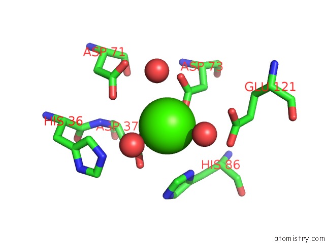
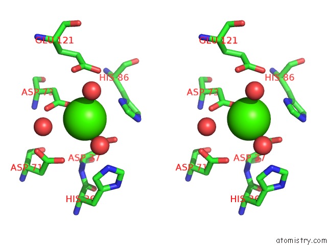
Calcium binding site 1 out of 4 in 1t6m
Go back to
Calcium binding site 1 out
of 4 in the X-Ray Structure of the R70D Pi-Plc Enzyme: Insight Into the Role of Calcium and Surrounding Amino Acids on Active Site Geometry and Catalysis.
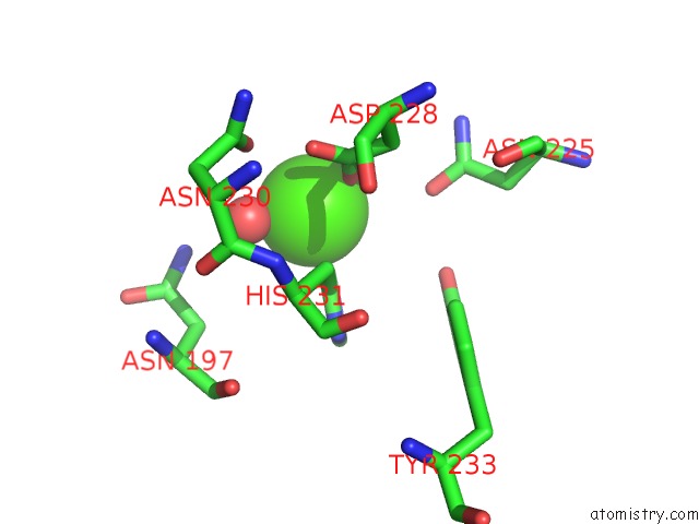
Mono view
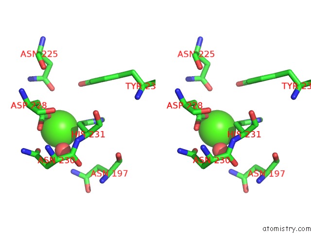
Stereo pair view
Mono view
Stereo pair view
A full contact list of Calcium with other atoms in the Ca binding
site number 1 of X-Ray Structure of the R70D Pi-Plc Enzyme: Insight Into the Role of Calcium and Surrounding Amino Acids on Active Site Geometry and Catalysis. within 5.0Å range:
|
Calcium binding site 2 out of 4 in 1t6m
Go back to
Calcium binding site 2 out
of 4 in the X-Ray Structure of the R70D Pi-Plc Enzyme: Insight Into the Role of Calcium and Surrounding Amino Acids on Active Site Geometry and Catalysis.
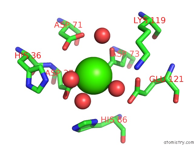
Mono view
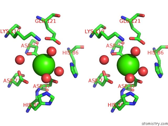
Stereo pair view
Mono view
Stereo pair view
A full contact list of Calcium with other atoms in the Ca binding
site number 2 of X-Ray Structure of the R70D Pi-Plc Enzyme: Insight Into the Role of Calcium and Surrounding Amino Acids on Active Site Geometry and Catalysis. within 5.0Å range:
|
Calcium binding site 3 out of 4 in 1t6m
Go back to
Calcium binding site 3 out
of 4 in the X-Ray Structure of the R70D Pi-Plc Enzyme: Insight Into the Role of Calcium and Surrounding Amino Acids on Active Site Geometry and Catalysis.
Mono view
Stereo pair view
Mono view
Stereo pair view
A full contact list of Calcium with other atoms in the Ca binding
site number 3 of X-Ray Structure of the R70D Pi-Plc Enzyme: Insight Into the Role of Calcium and Surrounding Amino Acids on Active Site Geometry and Catalysis. within 5.0Å range:
|
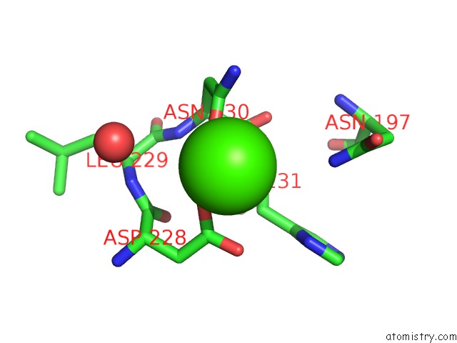
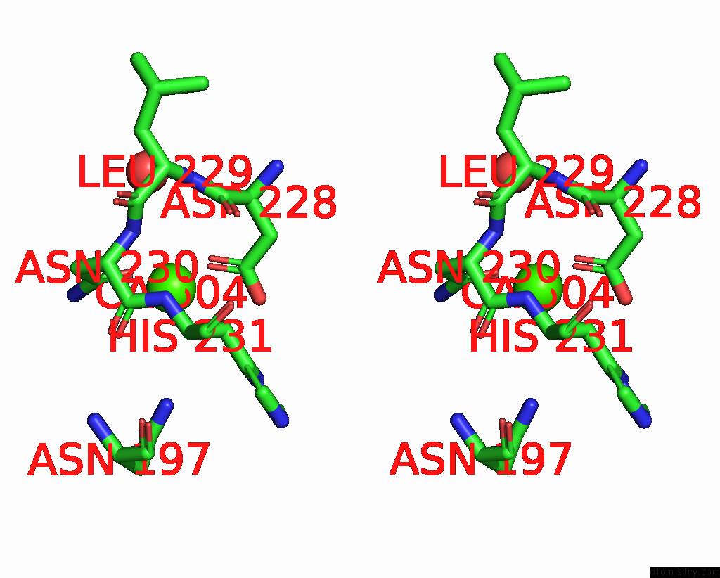
Calcium binding site 4 out of 4 in 1t6m
Go back to
Calcium binding site 4 out
of 4 in the X-Ray Structure of the R70D Pi-Plc Enzyme: Insight Into the Role of Calcium and Surrounding Amino Acids on Active Site Geometry and Catalysis.
Mono view
Stereo pair view
Mono view
Stereo pair view
A full contact list of Calcium with other atoms in the Ca binding
site number 4 of X-Ray Structure of the R70D Pi-Plc Enzyme: Insight Into the Role of Calcium and Surrounding Amino Acids on Active Site Geometry and Catalysis. within 5.0Å range:
|
Reference:
D.Apiyo,
L.Zhao,
M.-D.Tsai,
T.L.Selby.
X-Ray Structure of the R69D Phosphatidylinositol-Specific Phospholipase C Enzyme: Insight Into the Role of Calcium and Surrounding Amino Acids in Active Site Geometry and Catalysis. Biochemistry V. 44 9980 2005.
ISSN: ISSN 0006-2960
PubMed: 16042375
DOI: 10.1021/BI047896V
Page generated: Thu Jul 11 22:57:24 2024
ISSN: ISSN 0006-2960
PubMed: 16042375
DOI: 10.1021/BI047896V
Last articles
Zn in 9J0NZn in 9J0O
Zn in 9J0P
Zn in 9FJX
Zn in 9EKB
Zn in 9C0F
Zn in 9CAH
Zn in 9CH0
Zn in 9CH3
Zn in 9CH1