Calcium »
PDB 1t5t-1tkh »
1tgb »
Calcium in PDB 1tgb: Crystal Structure of Bovine Trypsinogen at 1.8 Angstroms Resolution. II. Crystallographic Refinement, Refined Crystal Structure and Comparison with Bovine Trypsin
Protein crystallography data
The structure of Crystal Structure of Bovine Trypsinogen at 1.8 Angstroms Resolution. II. Crystallographic Refinement, Refined Crystal Structure and Comparison with Bovine Trypsin, PDB code: 1tgb
was solved by
W.Bode,
H.Fehlhammer,
R.Huber,
with X-Ray Crystallography technique. A brief refinement statistics is given in the table below:
| Resolution Low / High (Å) | N/A / 1.80 |
| Space group | P 31 2 1 |
| Cell size a, b, c (Å), α, β, γ (°) | 55.100, 55.100, 109.400, 90.00, 90.00, 120.00 |
| R / Rfree (%) | n/a / n/a |
Calcium Binding Sites:
The binding sites of Calcium atom in the Crystal Structure of Bovine Trypsinogen at 1.8 Angstroms Resolution. II. Crystallographic Refinement, Refined Crystal Structure and Comparison with Bovine Trypsin
(pdb code 1tgb). This binding sites where shown within
5.0 Angstroms radius around Calcium atom.
In total only one binding site of Calcium was determined in the Crystal Structure of Bovine Trypsinogen at 1.8 Angstroms Resolution. II. Crystallographic Refinement, Refined Crystal Structure and Comparison with Bovine Trypsin, PDB code: 1tgb:
In total only one binding site of Calcium was determined in the Crystal Structure of Bovine Trypsinogen at 1.8 Angstroms Resolution. II. Crystallographic Refinement, Refined Crystal Structure and Comparison with Bovine Trypsin, PDB code: 1tgb:
Calcium binding site 1 out of 1 in 1tgb
Go back to
Calcium binding site 1 out
of 1 in the Crystal Structure of Bovine Trypsinogen at 1.8 Angstroms Resolution. II. Crystallographic Refinement, Refined Crystal Structure and Comparison with Bovine Trypsin
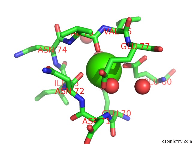
Mono view
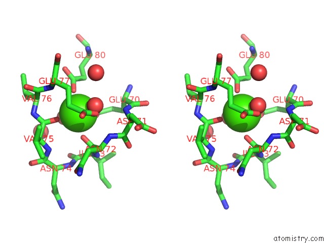
Stereo pair view
Mono view
Stereo pair view
A full contact list of Calcium with other atoms in the Ca binding
site number 1 of Crystal Structure of Bovine Trypsinogen at 1.8 Angstroms Resolution. II. Crystallographic Refinement, Refined Crystal Structure and Comparison with Bovine Trypsin within 5.0Å range:
|
Reference:
H.Fehlhammer,
W.Bode,
R.Huber.
Crystal Structure of Bovine Trypsinogen at 1-8 A Resolution. II. Crystallographic Refinement, Refined Crystal Structure and Comparison with Bovine Trypsin. J.Mol.Biol. V. 111 415 1977.
ISSN: ISSN 0022-2836
PubMed: 864704
DOI: 10.1016/S0022-2836(77)80062-4
Page generated: Thu Jul 11 23:02:15 2024
ISSN: ISSN 0022-2836
PubMed: 864704
DOI: 10.1016/S0022-2836(77)80062-4
Last articles
Zn in 9J0NZn in 9J0O
Zn in 9J0P
Zn in 9FJX
Zn in 9EKB
Zn in 9C0F
Zn in 9CAH
Zn in 9CH0
Zn in 9CH3
Zn in 9CH1