Calcium »
PDB 1tu5-1ujc »
1tu5 »
Calcium in PDB 1tu5: Crystal Structure of Bovine Plasma Copper-Containing Amine Oxidase
Enzymatic activity of Crystal Structure of Bovine Plasma Copper-Containing Amine Oxidase
All present enzymatic activity of Crystal Structure of Bovine Plasma Copper-Containing Amine Oxidase:
1.4.3.6;
1.4.3.6;
Protein crystallography data
The structure of Crystal Structure of Bovine Plasma Copper-Containing Amine Oxidase, PDB code: 1tu5
was solved by
M.Lunelli,
M.L.Di Paolo,
M.Biadene,
V.Calderone,
M.Scarpa,
R.Battistutta,
A.Rigo,
G.Zanotti,
with X-Ray Crystallography technique. A brief refinement statistics is given in the table below:
| Resolution Low / High (Å) | 25.00 / 2.37 |
| Space group | P 21 21 21 |
| Cell size a, b, c (Å), α, β, γ (°) | 77.680, 131.192, 134.001, 90.00, 90.00, 90.00 |
| R / Rfree (%) | 20.5 / 23.7 |
Other elements in 1tu5:
The structure of Crystal Structure of Bovine Plasma Copper-Containing Amine Oxidase also contains other interesting chemical elements:
| Copper | (Cu) | 2 atoms |
| Chlorine | (Cl) | 3 atoms |
Calcium Binding Sites:
The binding sites of Calcium atom in the Crystal Structure of Bovine Plasma Copper-Containing Amine Oxidase
(pdb code 1tu5). This binding sites where shown within
5.0 Angstroms radius around Calcium atom.
In total 4 binding sites of Calcium where determined in the Crystal Structure of Bovine Plasma Copper-Containing Amine Oxidase, PDB code: 1tu5:
Jump to Calcium binding site number: 1; 2; 3; 4;
In total 4 binding sites of Calcium where determined in the Crystal Structure of Bovine Plasma Copper-Containing Amine Oxidase, PDB code: 1tu5:
Jump to Calcium binding site number: 1; 2; 3; 4;
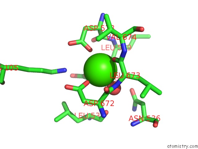
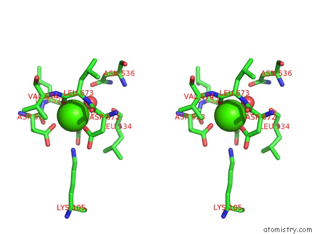
Calcium binding site 1 out of 4 in 1tu5
Go back to
Calcium binding site 1 out
of 4 in the Crystal Structure of Bovine Plasma Copper-Containing Amine Oxidase
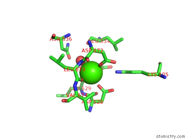
Mono view
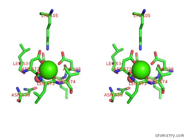
Stereo pair view
Mono view
Stereo pair view
A full contact list of Calcium with other atoms in the Ca binding
site number 1 of Crystal Structure of Bovine Plasma Copper-Containing Amine Oxidase within 5.0Å range:
|
Calcium binding site 2 out of 4 in 1tu5
Go back to
Calcium binding site 2 out
of 4 in the Crystal Structure of Bovine Plasma Copper-Containing Amine Oxidase
Mono view
Stereo pair view
Mono view
Stereo pair view
A full contact list of Calcium with other atoms in the Ca binding
site number 2 of Crystal Structure of Bovine Plasma Copper-Containing Amine Oxidase within 5.0Å range:
|
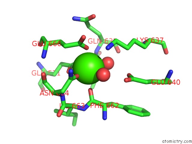
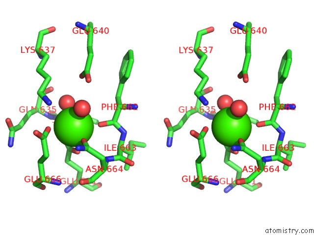
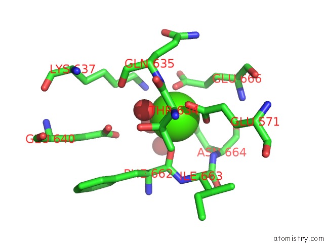
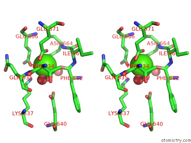
Calcium binding site 3 out of 4 in 1tu5
Go back to
Calcium binding site 3 out
of 4 in the Crystal Structure of Bovine Plasma Copper-Containing Amine Oxidase
Mono view
Stereo pair view
Mono view
Stereo pair view
A full contact list of Calcium with other atoms in the Ca binding
site number 3 of Crystal Structure of Bovine Plasma Copper-Containing Amine Oxidase within 5.0Å range:
|
Calcium binding site 4 out of 4 in 1tu5
Go back to
Calcium binding site 4 out
of 4 in the Crystal Structure of Bovine Plasma Copper-Containing Amine Oxidase
Mono view
Stereo pair view
Mono view
Stereo pair view
A full contact list of Calcium with other atoms in the Ca binding
site number 4 of Crystal Structure of Bovine Plasma Copper-Containing Amine Oxidase within 5.0Å range:
|
Reference:
M.Lunelli,
M.L.Di Paolo,
M.Biadene,
V.Calderone,
R.Battistutta,
M.Scarpa,
A.Rigo,
G.Zanotti.
Crystal Structure of Amine Oxidase From Bovine Serum. J.Mol.Biol. V. 346 991 2005.
ISSN: ISSN 0022-2836
PubMed: 15701511
DOI: 10.1016/J.JMB.2004.12.038
Page generated: Tue Jul 8 02:21:56 2025
ISSN: ISSN 0022-2836
PubMed: 15701511
DOI: 10.1016/J.JMB.2004.12.038
Last articles
Fe in 2YXOFe in 2YRS
Fe in 2YXC
Fe in 2YNM
Fe in 2YVJ
Fe in 2YP1
Fe in 2YU2
Fe in 2YU1
Fe in 2YQB
Fe in 2YOO