Calcium »
PDB 1tu5-1ujc »
1u5q »
Calcium in PDB 1u5q: Crystal Structure of the TAO2 Kinase Domain: Activation and Specifity of A STE20P MAP3K
Protein crystallography data
The structure of Crystal Structure of the TAO2 Kinase Domain: Activation and Specifity of A STE20P MAP3K, PDB code: 1u5q
was solved by
T.Zhou,
M.Raman,
Y.Gao,
S.Earnest,
Z.Chen,
M.Machius,
M.H.Cobb,
E.J.Goldsmith,
with X-Ray Crystallography technique. A brief refinement statistics is given in the table below:
| Resolution Low / High (Å) | 50.00 / 2.10 |
| Space group | P 65 2 2 |
| Cell size a, b, c (Å), α, β, γ (°) | 186.220, 186.220, 94.506, 90.00, 90.00, 120.00 |
| R / Rfree (%) | 22.3 / 26.9 |
Calcium Binding Sites:
The binding sites of Calcium atom in the Crystal Structure of the TAO2 Kinase Domain: Activation and Specifity of A STE20P MAP3K
(pdb code 1u5q). This binding sites where shown within
5.0 Angstroms radius around Calcium atom.
In total 2 binding sites of Calcium where determined in the Crystal Structure of the TAO2 Kinase Domain: Activation and Specifity of A STE20P MAP3K, PDB code: 1u5q:
Jump to Calcium binding site number: 1; 2;
In total 2 binding sites of Calcium where determined in the Crystal Structure of the TAO2 Kinase Domain: Activation and Specifity of A STE20P MAP3K, PDB code: 1u5q:
Jump to Calcium binding site number: 1; 2;
Calcium binding site 1 out of 2 in 1u5q
Go back to
Calcium binding site 1 out
of 2 in the Crystal Structure of the TAO2 Kinase Domain: Activation and Specifity of A STE20P MAP3K
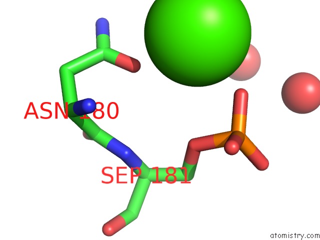
Mono view
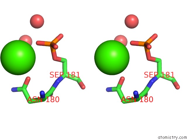
Stereo pair view
Mono view
Stereo pair view
A full contact list of Calcium with other atoms in the Ca binding
site number 1 of Crystal Structure of the TAO2 Kinase Domain: Activation and Specifity of A STE20P MAP3K within 5.0Å range:
|
Calcium binding site 2 out of 2 in 1u5q
Go back to
Calcium binding site 2 out
of 2 in the Crystal Structure of the TAO2 Kinase Domain: Activation and Specifity of A STE20P MAP3K
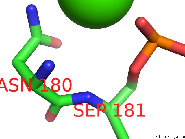
Mono view
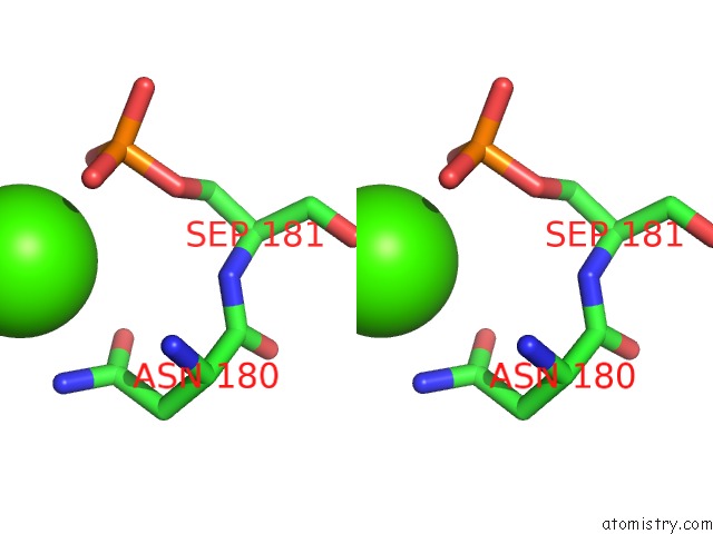
Stereo pair view
Mono view
Stereo pair view
A full contact list of Calcium with other atoms in the Ca binding
site number 2 of Crystal Structure of the TAO2 Kinase Domain: Activation and Specifity of A STE20P MAP3K within 5.0Å range:
|
Reference:
T.Zhou,
M.Raman,
Y.Gao,
S.Earnest,
Z.Chen,
M.Machius,
M.H.Cobb,
E.J.Goldsmith.
Crystal Structure of the TAO2 Kinase Domain; Activation and Specificity of A STE20P MAP3K. Structure V. 12 1891 2004.
ISSN: ISSN 0969-2126
PubMed: 15458637
DOI: 10.1016/J.STR.2004.07.021
Page generated: Thu Jul 11 23:16:33 2024
ISSN: ISSN 0969-2126
PubMed: 15458637
DOI: 10.1016/J.STR.2004.07.021
Last articles
Zn in 9MJ5Zn in 9HNW
Zn in 9G0L
Zn in 9FNE
Zn in 9DZN
Zn in 9E0I
Zn in 9D32
Zn in 9DAK
Zn in 8ZXC
Zn in 8ZUF