Calcium »
PDB 1tu5-1ujc »
1ua7 »
Calcium in PDB 1ua7: Crystal Structure Analysis of Alpha-Amylase From Bacillus Subtilis Complexed with Acarbose
Enzymatic activity of Crystal Structure Analysis of Alpha-Amylase From Bacillus Subtilis Complexed with Acarbose
All present enzymatic activity of Crystal Structure Analysis of Alpha-Amylase From Bacillus Subtilis Complexed with Acarbose:
3.2.1.1;
3.2.1.1;
Protein crystallography data
The structure of Crystal Structure Analysis of Alpha-Amylase From Bacillus Subtilis Complexed with Acarbose, PDB code: 1ua7
was solved by
M.Kagawa,
Z.Fujimoto,
M.Momma,
K.Takase,
H.Mizuno,
with X-Ray Crystallography technique. A brief refinement statistics is given in the table below:
| Resolution Low / High (Å) | 19.97 / 2.21 |
| Space group | P 21 21 21 |
| Cell size a, b, c (Å), α, β, γ (°) | 70.320, 74.204, 115.724, 90.00, 90.00, 90.00 |
| R / Rfree (%) | 20.8 / 26.5 |
Calcium Binding Sites:
The binding sites of Calcium atom in the Crystal Structure Analysis of Alpha-Amylase From Bacillus Subtilis Complexed with Acarbose
(pdb code 1ua7). This binding sites where shown within
5.0 Angstroms radius around Calcium atom.
In total 3 binding sites of Calcium where determined in the Crystal Structure Analysis of Alpha-Amylase From Bacillus Subtilis Complexed with Acarbose, PDB code: 1ua7:
Jump to Calcium binding site number: 1; 2; 3;
In total 3 binding sites of Calcium where determined in the Crystal Structure Analysis of Alpha-Amylase From Bacillus Subtilis Complexed with Acarbose, PDB code: 1ua7:
Jump to Calcium binding site number: 1; 2; 3;
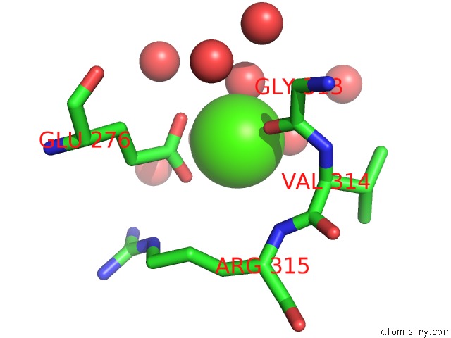
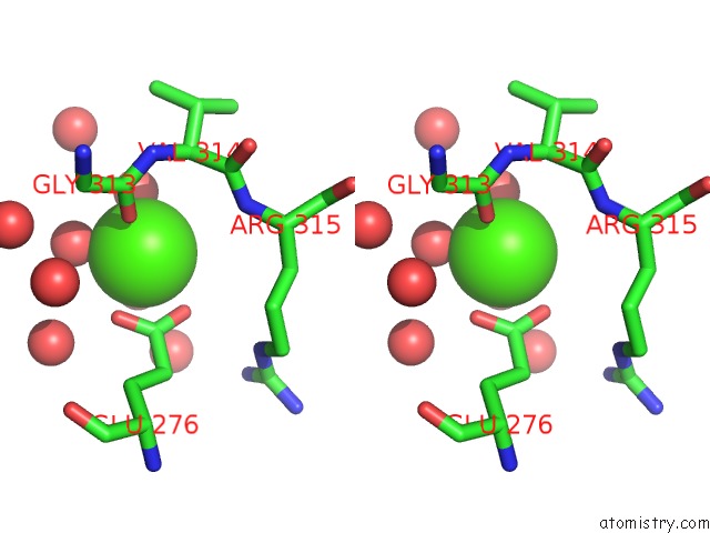
Calcium binding site 1 out of 3 in 1ua7
Go back to
Calcium binding site 1 out
of 3 in the Crystal Structure Analysis of Alpha-Amylase From Bacillus Subtilis Complexed with Acarbose
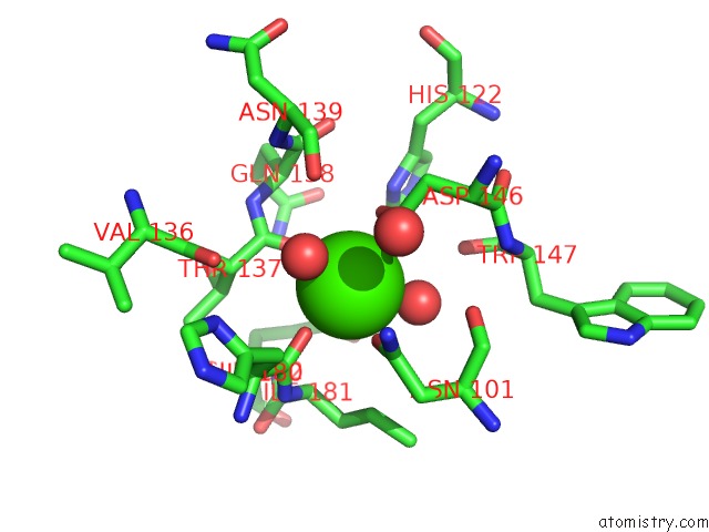
Mono view
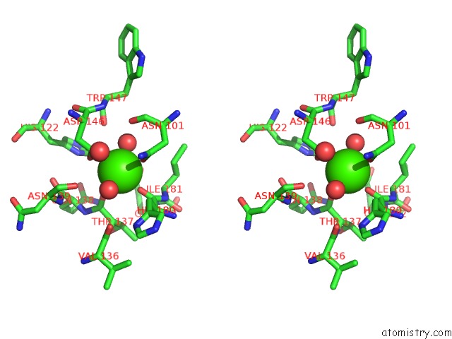
Stereo pair view
Mono view
Stereo pair view
A full contact list of Calcium with other atoms in the Ca binding
site number 1 of Crystal Structure Analysis of Alpha-Amylase From Bacillus Subtilis Complexed with Acarbose within 5.0Å range:
|
Calcium binding site 2 out of 3 in 1ua7
Go back to
Calcium binding site 2 out
of 3 in the Crystal Structure Analysis of Alpha-Amylase From Bacillus Subtilis Complexed with Acarbose
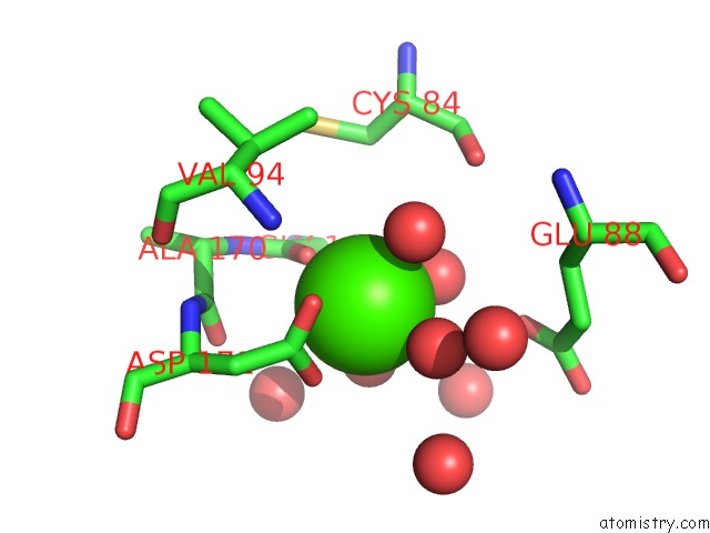
Mono view
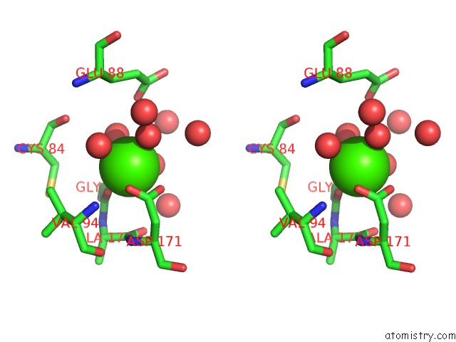
Stereo pair view
Mono view
Stereo pair view
A full contact list of Calcium with other atoms in the Ca binding
site number 2 of Crystal Structure Analysis of Alpha-Amylase From Bacillus Subtilis Complexed with Acarbose within 5.0Å range:
|
Calcium binding site 3 out of 3 in 1ua7
Go back to
Calcium binding site 3 out
of 3 in the Crystal Structure Analysis of Alpha-Amylase From Bacillus Subtilis Complexed with Acarbose
Mono view
Stereo pair view
Mono view
Stereo pair view
A full contact list of Calcium with other atoms in the Ca binding
site number 3 of Crystal Structure Analysis of Alpha-Amylase From Bacillus Subtilis Complexed with Acarbose within 5.0Å range:
|
Reference:
M.Kagawa,
Z.Fujimoto,
M.Momma,
K.Takase,
H.Mizuno.
Crystal Structure of Bacillus Subtilis Alpha-Amylase in Complex with Acarbose J.Bacteriol. V. 185 6981 2003.
ISSN: ISSN 0021-9193
PubMed: 14617662
DOI: 10.1128/JB.185.23.6981-6984.2003
Page generated: Thu Jul 11 23:17:49 2024
ISSN: ISSN 0021-9193
PubMed: 14617662
DOI: 10.1128/JB.185.23.6981-6984.2003
Last articles
Zn in 9J0NZn in 9J0O
Zn in 9J0P
Zn in 9FJX
Zn in 9EKB
Zn in 9C0F
Zn in 9CAH
Zn in 9CH0
Zn in 9CH3
Zn in 9CH1