Calcium »
PDB 1tu5-1ujc »
1ucn »
Calcium in PDB 1ucn: X-Ray Structure of Human Nucleoside Diphosphate Kinase A Complexed with Adp at 2 A Resolution
Enzymatic activity of X-Ray Structure of Human Nucleoside Diphosphate Kinase A Complexed with Adp at 2 A Resolution
All present enzymatic activity of X-Ray Structure of Human Nucleoside Diphosphate Kinase A Complexed with Adp at 2 A Resolution:
2.7.4.6;
2.7.4.6;
Protein crystallography data
The structure of X-Ray Structure of Human Nucleoside Diphosphate Kinase A Complexed with Adp at 2 A Resolution, PDB code: 1ucn
was solved by
Y.Chen,
S.Gallois-Montbrun,
B.Schneider,
M.Veron,
S.Morera,
D.Deville-Bonne,
J.Janin,
with X-Ray Crystallography technique. A brief refinement statistics is given in the table below:
| Resolution Low / High (Å) | 19.95 / 2.00 |
| Space group | P 41 21 2 |
| Cell size a, b, c (Å), α, β, γ (°) | 116.075, 116.075, 91.292, 90.00, 90.00, 90.00 |
| R / Rfree (%) | 23.7 / 28.2 |
Calcium Binding Sites:
The binding sites of Calcium atom in the X-Ray Structure of Human Nucleoside Diphosphate Kinase A Complexed with Adp at 2 A Resolution
(pdb code 1ucn). This binding sites where shown within
5.0 Angstroms radius around Calcium atom.
In total 5 binding sites of Calcium where determined in the X-Ray Structure of Human Nucleoside Diphosphate Kinase A Complexed with Adp at 2 A Resolution, PDB code: 1ucn:
Jump to Calcium binding site number: 1; 2; 3; 4; 5;
In total 5 binding sites of Calcium where determined in the X-Ray Structure of Human Nucleoside Diphosphate Kinase A Complexed with Adp at 2 A Resolution, PDB code: 1ucn:
Jump to Calcium binding site number: 1; 2; 3; 4; 5;
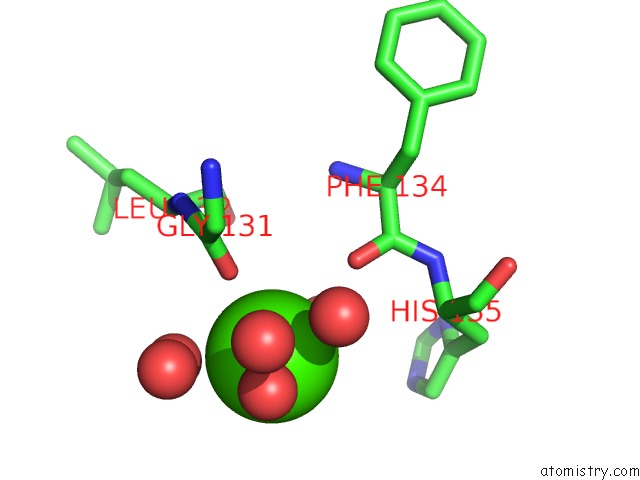
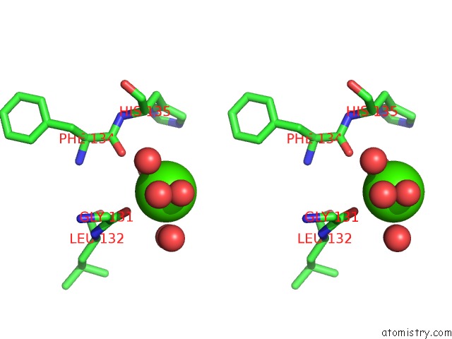
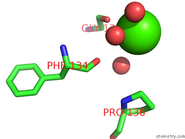
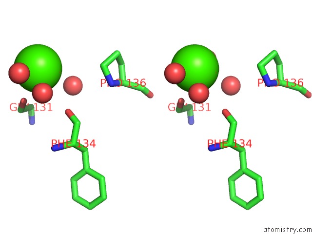
Calcium binding site 1 out of 5 in 1ucn
Go back to
Calcium binding site 1 out
of 5 in the X-Ray Structure of Human Nucleoside Diphosphate Kinase A Complexed with Adp at 2 A Resolution
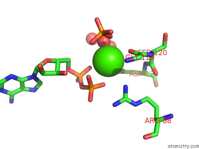
Mono view
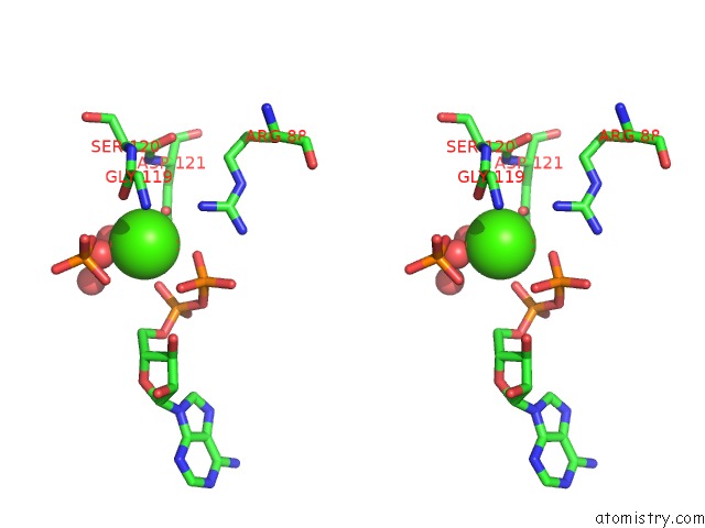
Stereo pair view
Mono view
Stereo pair view
A full contact list of Calcium with other atoms in the Ca binding
site number 1 of X-Ray Structure of Human Nucleoside Diphosphate Kinase A Complexed with Adp at 2 A Resolution within 5.0Å range:
|
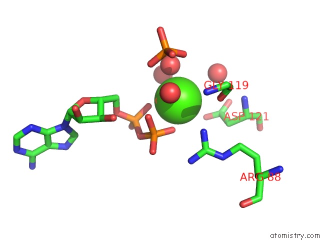
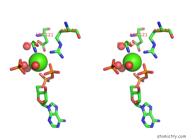
Calcium binding site 2 out of 5 in 1ucn
Go back to
Calcium binding site 2 out
of 5 in the X-Ray Structure of Human Nucleoside Diphosphate Kinase A Complexed with Adp at 2 A Resolution
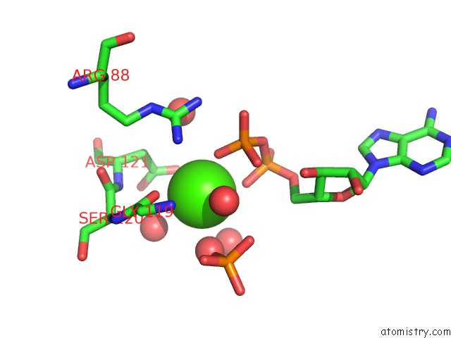
Mono view
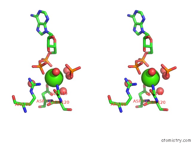
Stereo pair view
Mono view
Stereo pair view
A full contact list of Calcium with other atoms in the Ca binding
site number 2 of X-Ray Structure of Human Nucleoside Diphosphate Kinase A Complexed with Adp at 2 A Resolution within 5.0Å range:
|
Calcium binding site 3 out of 5 in 1ucn
Go back to
Calcium binding site 3 out
of 5 in the X-Ray Structure of Human Nucleoside Diphosphate Kinase A Complexed with Adp at 2 A Resolution
Mono view
Stereo pair view
Mono view
Stereo pair view
A full contact list of Calcium with other atoms in the Ca binding
site number 3 of X-Ray Structure of Human Nucleoside Diphosphate Kinase A Complexed with Adp at 2 A Resolution within 5.0Å range:
|
Calcium binding site 4 out of 5 in 1ucn
Go back to
Calcium binding site 4 out
of 5 in the X-Ray Structure of Human Nucleoside Diphosphate Kinase A Complexed with Adp at 2 A Resolution
Mono view
Stereo pair view
Mono view
Stereo pair view
A full contact list of Calcium with other atoms in the Ca binding
site number 4 of X-Ray Structure of Human Nucleoside Diphosphate Kinase A Complexed with Adp at 2 A Resolution within 5.0Å range:
|
Calcium binding site 5 out of 5 in 1ucn
Go back to
Calcium binding site 5 out
of 5 in the X-Ray Structure of Human Nucleoside Diphosphate Kinase A Complexed with Adp at 2 A Resolution
Mono view
Stereo pair view
Mono view
Stereo pair view
A full contact list of Calcium with other atoms in the Ca binding
site number 5 of X-Ray Structure of Human Nucleoside Diphosphate Kinase A Complexed with Adp at 2 A Resolution within 5.0Å range:
|
Reference:
Y.Chen,
S.Gallois-Montbrun,
B.Schneider,
M.Veron,
S.Morera,
D.Deville-Bonne,
J.Janin.
Nucleotide Binding to Nucleoside Diphosphate Kinases: X-Ray Structure of Human Ndpk-A in Complex with Adp and Comparison to Protein Kinases J.Mol.Biol. V. 332 915 2003.
ISSN: ISSN 0022-2836
PubMed: 12972261
DOI: 10.1016/J.JMB.2003.07.004
Page generated: Thu Jul 11 23:18:31 2024
ISSN: ISSN 0022-2836
PubMed: 12972261
DOI: 10.1016/J.JMB.2003.07.004
Last articles
Zn in 9J0NZn in 9J0O
Zn in 9J0P
Zn in 9FJX
Zn in 9EKB
Zn in 9C0F
Zn in 9CAH
Zn in 9CH0
Zn in 9CH3
Zn in 9CH1