Calcium »
PDB 1ukg-1ux6 »
1ukq »
Calcium in PDB 1ukq: Crystal Structure of Cyclodextrin Glucanotransferase Complexed with A Pseudo-Maltotetraose Derived From Acarbose
Enzymatic activity of Crystal Structure of Cyclodextrin Glucanotransferase Complexed with A Pseudo-Maltotetraose Derived From Acarbose
All present enzymatic activity of Crystal Structure of Cyclodextrin Glucanotransferase Complexed with A Pseudo-Maltotetraose Derived From Acarbose:
2.4.1.19;
2.4.1.19;
Protein crystallography data
The structure of Crystal Structure of Cyclodextrin Glucanotransferase Complexed with A Pseudo-Maltotetraose Derived From Acarbose, PDB code: 1ukq
was solved by
K.Haga,
R.Kanai,
O.Sakamoto,
K.Harata,
K.Yamane,
with X-Ray Crystallography technique. A brief refinement statistics is given in the table below:
| Resolution Low / High (Å) | 10.00 / 2.00 |
| Space group | P 1 |
| Cell size a, b, c (Å), α, β, γ (°) | 64.940, 74.490, 79.100, 85.13, 105.02, 101.02 |
| R / Rfree (%) | 18.4 / 24.6 |
Calcium Binding Sites:
The binding sites of Calcium atom in the Crystal Structure of Cyclodextrin Glucanotransferase Complexed with A Pseudo-Maltotetraose Derived From Acarbose
(pdb code 1ukq). This binding sites where shown within
5.0 Angstroms radius around Calcium atom.
In total 4 binding sites of Calcium where determined in the Crystal Structure of Cyclodextrin Glucanotransferase Complexed with A Pseudo-Maltotetraose Derived From Acarbose, PDB code: 1ukq:
Jump to Calcium binding site number: 1; 2; 3; 4;
In total 4 binding sites of Calcium where determined in the Crystal Structure of Cyclodextrin Glucanotransferase Complexed with A Pseudo-Maltotetraose Derived From Acarbose, PDB code: 1ukq:
Jump to Calcium binding site number: 1; 2; 3; 4;
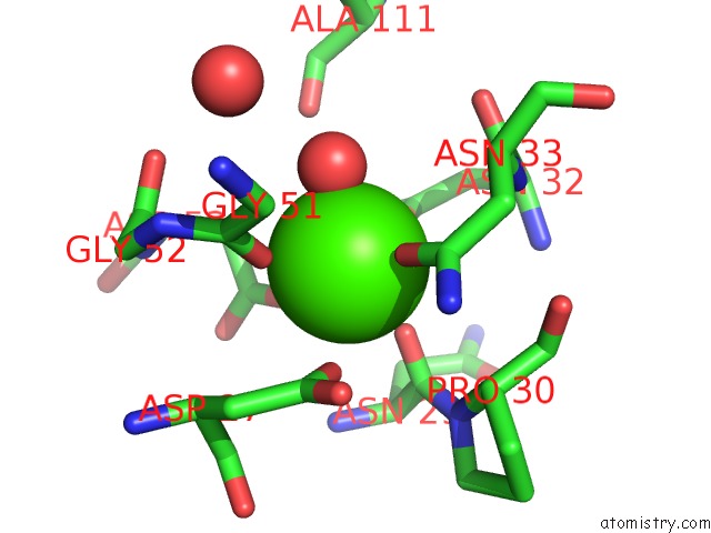
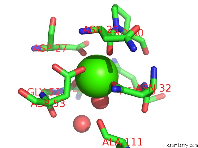
Calcium binding site 1 out of 4 in 1ukq
Go back to
Calcium binding site 1 out
of 4 in the Crystal Structure of Cyclodextrin Glucanotransferase Complexed with A Pseudo-Maltotetraose Derived From Acarbose
Mono view
Stereo pair view
Mono view
Stereo pair view
A full contact list of Calcium with other atoms in the Ca binding
site number 1 of Crystal Structure of Cyclodextrin Glucanotransferase Complexed with A Pseudo-Maltotetraose Derived From Acarbose within 5.0Å range:
|
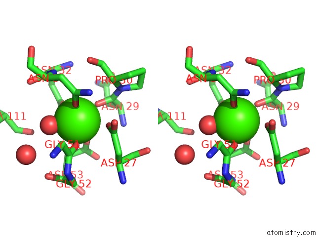
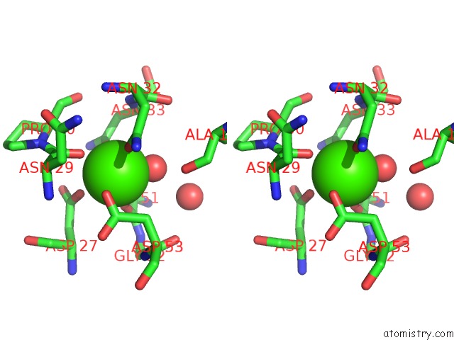
Calcium binding site 2 out of 4 in 1ukq
Go back to
Calcium binding site 2 out
of 4 in the Crystal Structure of Cyclodextrin Glucanotransferase Complexed with A Pseudo-Maltotetraose Derived From Acarbose
Mono view
Stereo pair view
Mono view
Stereo pair view
A full contact list of Calcium with other atoms in the Ca binding
site number 2 of Crystal Structure of Cyclodextrin Glucanotransferase Complexed with A Pseudo-Maltotetraose Derived From Acarbose within 5.0Å range:
|
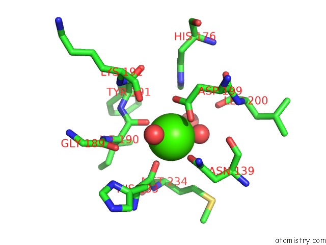
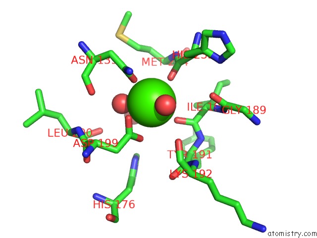
Calcium binding site 3 out of 4 in 1ukq
Go back to
Calcium binding site 3 out
of 4 in the Crystal Structure of Cyclodextrin Glucanotransferase Complexed with A Pseudo-Maltotetraose Derived From Acarbose
Mono view
Stereo pair view
Mono view
Stereo pair view
A full contact list of Calcium with other atoms in the Ca binding
site number 3 of Crystal Structure of Cyclodextrin Glucanotransferase Complexed with A Pseudo-Maltotetraose Derived From Acarbose within 5.0Å range:
|
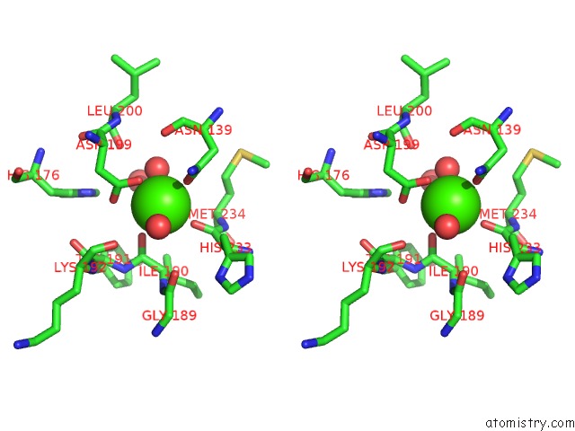
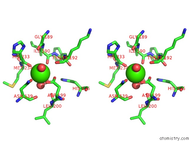
Calcium binding site 4 out of 4 in 1ukq
Go back to
Calcium binding site 4 out
of 4 in the Crystal Structure of Cyclodextrin Glucanotransferase Complexed with A Pseudo-Maltotetraose Derived From Acarbose
Mono view
Stereo pair view
Mono view
Stereo pair view
A full contact list of Calcium with other atoms in the Ca binding
site number 4 of Crystal Structure of Cyclodextrin Glucanotransferase Complexed with A Pseudo-Maltotetraose Derived From Acarbose within 5.0Å range:
|
Reference:
K.Haga,
R.Kanai,
O.Sakamoto,
M.Aoyagi,
K.Harata,
K.Yamane.
Effects of Essential Carbohydrate/Aromatic Stacking Interaction with TYR100 and PHE259 on Substrate Binding of Cyclodextrin Glycosyltransferase From Alkalophilic Bacillus Sp. 1011 J.Biochem.(Tokyo) V. 134 881 2003.
ISSN: ISSN 0021-924X
PubMed: 14769878
DOI: 10.1093/JB/MVG215
Page generated: Thu Jul 11 23:31:40 2024
ISSN: ISSN 0021-924X
PubMed: 14769878
DOI: 10.1093/JB/MVG215
Last articles
Zn in 9J0NZn in 9J0O
Zn in 9J0P
Zn in 9FJX
Zn in 9EKB
Zn in 9C0F
Zn in 9CAH
Zn in 9CH0
Zn in 9CH3
Zn in 9CH1